Department of Pathophysiology and Allergy Research; Center for Pathophysiology, Infectiology, and Immunology, Medical University of Vienna, Vienna, Austria; Department of Environmental Health Sciences, Bloomberg School of Public Health, Johns Hopkins University, Baltimore, Maryland, United States of America.
Department of Pathophysiology and Allergy Research; Center for Pathophysiology, Infectiology, and Immunology, Medical University of Vienna, Vienna, Austria.
PLoS One. 2015 Jan 30;10(1):e0117904. doi: 10.1371/journal.pone.0117904. eCollection 2015.
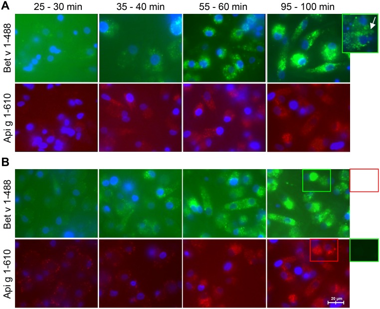
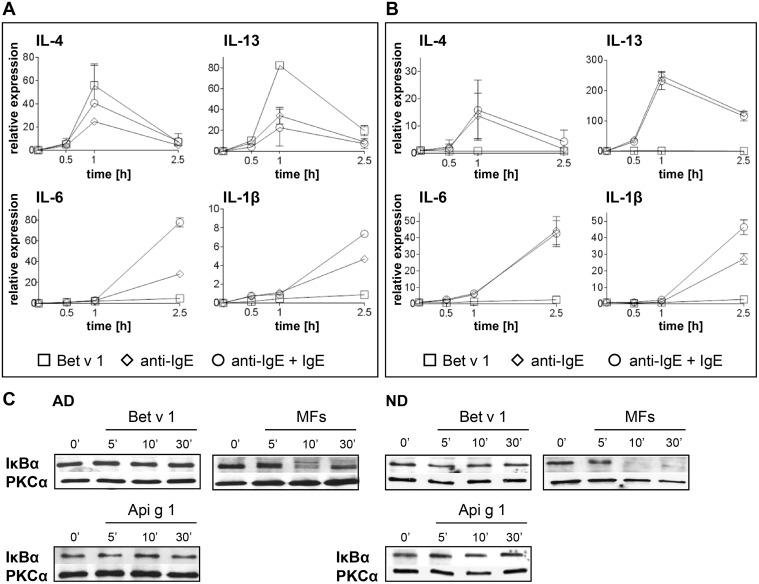
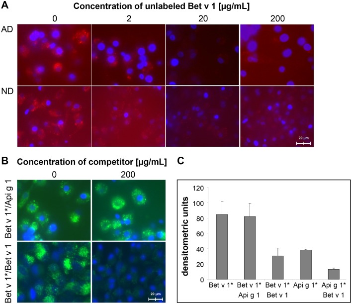
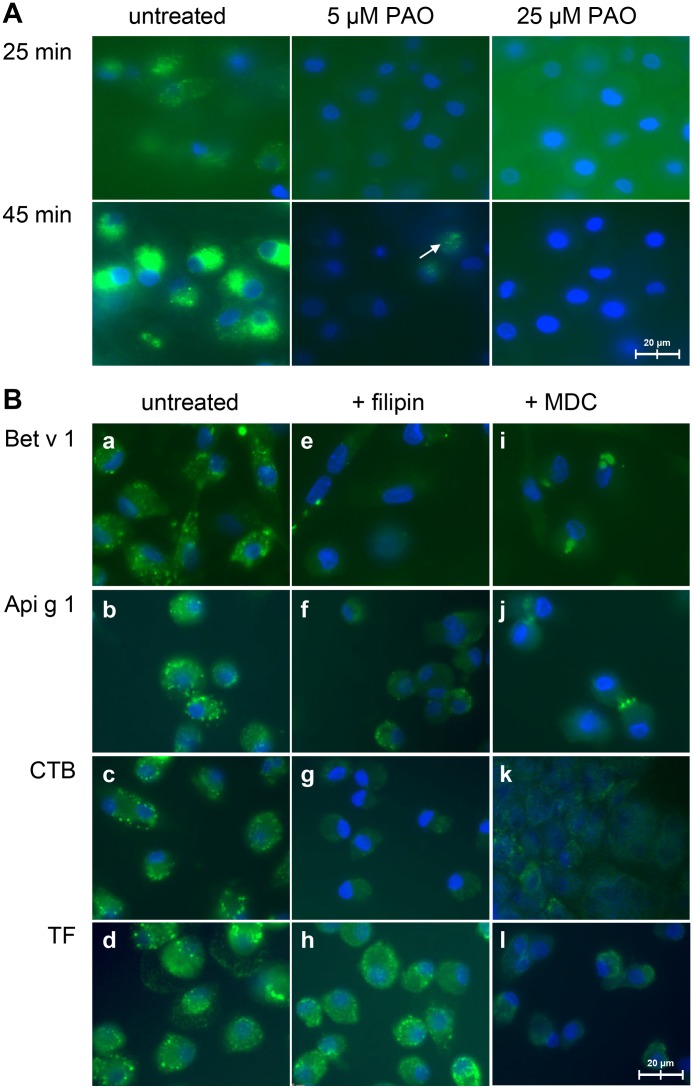
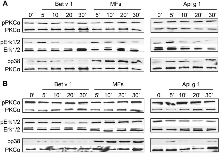
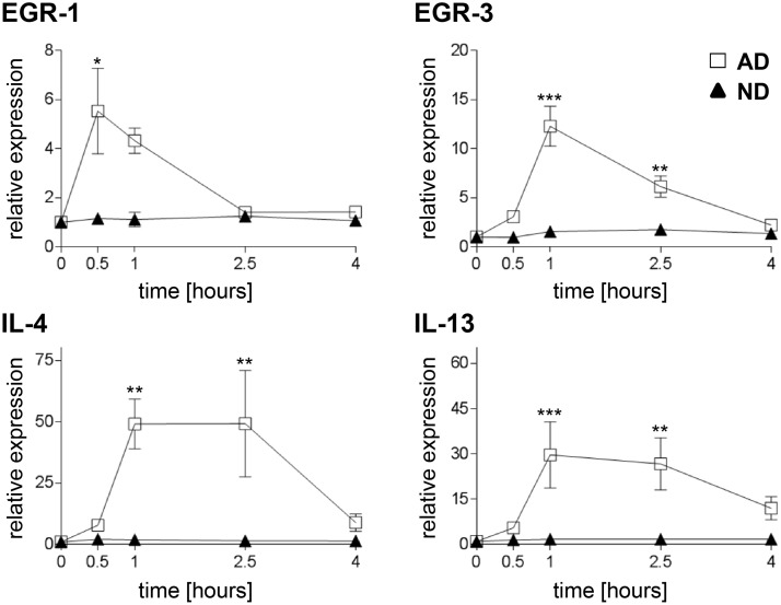
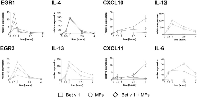
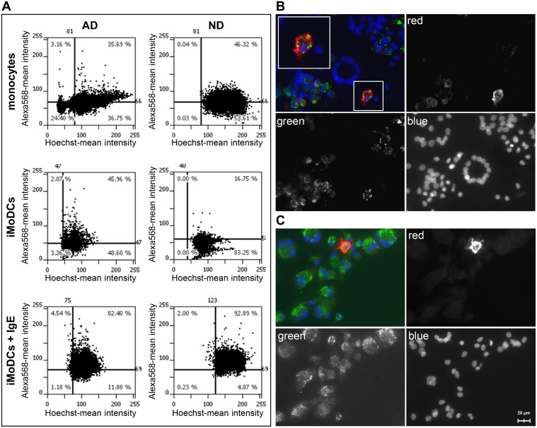
Dendritic cells play a fundamental role in shaping the immune response to allergens. The events that lead to allergic sensitization or tolerance induction during the interaction of the major birch pollen allergen Bet v 1 and dendritic cells are not very well studied. Here, we analyzed the uptake of Bet v 1 and the cross-reactive celery allergen Api g 1 by immature monocyte-derived dendritic cells (iMoDCs) of allergic and normal donors. In addition, we characterized the allergen-triggered intracellular signaling and transcriptional events. Uptake kinetics, competitive binding, and internalization pathways of labeled allergens by iMoDCs were visualized by live-cell imaging. Surface-bound IgE was detected by immunofluorescence microscopy and flow cytometry. Allergen- and IgE-induced gene expression of early growth response genes and Th1 and Th2 related cytokines and chemokines were analyzed by real-time PCR. Phosporylation of signaling kinases was analyzed by Western blot. Internalization of Bet v 1 by iMoDCs of both donor groups, likely by receptor-mediated caveolar endocytosis, followed similar kinetics. Bet v 1 outcompeted Api g 1 in cell surface binding and uptake. MoDCs of allergic and healthy donors displayed surface-bound IgE and showed a pronounced upregulation of Th2 cytokine- and NFκB-dependent genes upon non-specific Fcε receptor cross-linking. In contrast to these IgE-mediated responses, Bet v 1-stimulation increased transcript levels of the Th2 cytokines IL-4 and IL-13 but not of NFκB-related genes in MoDCs of BP allergic donors. Cells of healthy donors were either unresponsive or showed elevated mRNA levels of Th1-promoting chemokines. Moreover, Bet v 1 was able to induce Erk1/2 and p38 MAPK activation in BP allergics but only a slight p38 activation in normal donors. In conclusion, our data indicate that Bet v 1 favors the activation of a Th2 program only in DCs of BP allergic individuals.
树突状细胞在塑造对过敏原的免疫反应方面起着至关重要的作用。在主要桦树花粉过敏原 Bet v 1 与树突状细胞相互作用过程中导致过敏致敏或耐受诱导的事件尚未得到很好的研究。在这里,我们分析了过敏和正常供体的未成熟单核细胞衍生树突状细胞(iMoDC)对 Bet v 1 和交叉反应的芹菜过敏原 Api g 1 的摄取。此外,我们还对过敏原触发的细胞内信号和转录事件进行了表征。通过活细胞成像可视化 iMoDC 摄取标记过敏原的摄取动力学、竞争结合和内化途径。通过免疫荧光显微镜和流式细胞术检测表面结合的 IgE。通过实时 PCR 分析过敏原和 IgE 诱导的早期生长反应基因以及 Th1 和 Th2 相关细胞因子和趋化因子的基因表达。通过 Western blot 分析信号激酶的磷酸化。两组供体的 iMoDC 均能内化 Bet v 1,可能通过受体介导的小窝内吞作用,其内化动力学相似。Bet v 1 在细胞表面结合和摄取中竞争 Api g 1。过敏和健康供体的 MoDC 显示表面结合的 IgE,并在非特异性 Fcε受体交联时显示出 Th2 细胞因子和 NFκB 依赖性基因的显著上调。与这些 IgE 介导的反应相反,Bet v 1 刺激增加了 BP 过敏供体 MoDC 中 Th2 细胞因子 IL-4 和 IL-13 的转录水平,但不增加 NFκB 相关基因的转录水平。健康供体的细胞要么无反应,要么表现出 Th1 促进趋化因子的 mRNA 水平升高。此外,Bet v 1 能够在 BP 过敏个体的 DC 中诱导 Erk1/2 和 p38 MAPK 激活,但在正常供体中仅诱导轻微的 p38 激活。总之,我们的数据表明,Bet v 1 仅在 BP 过敏个体的 DC 中有利于激活 Th2 程序。