Rajaram Narasimhan, Reesor Andrew F, Mulvey Christine S, Frees Amy E, Ramanujam Nirmala
Department of Biomedical Engineering, Duke University, Durham, North Carolina, United States of America.
PLoS One. 2015 Jan 30;10(1):e0117132. doi: 10.1371/journal.pone.0117132. eCollection 2015.
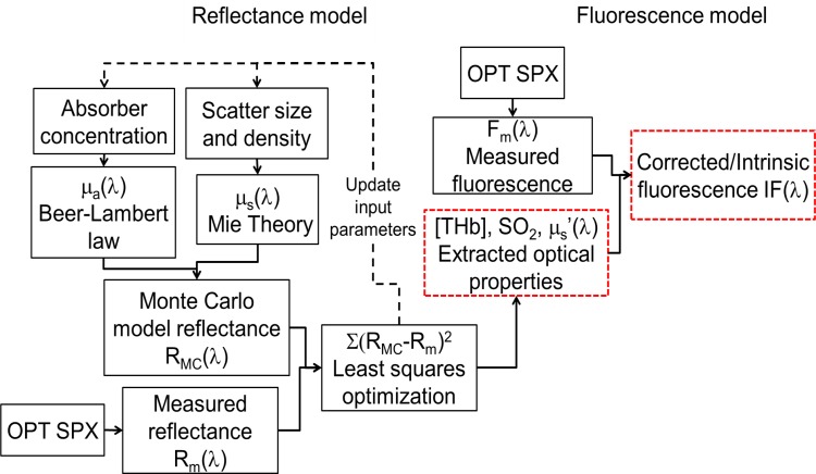
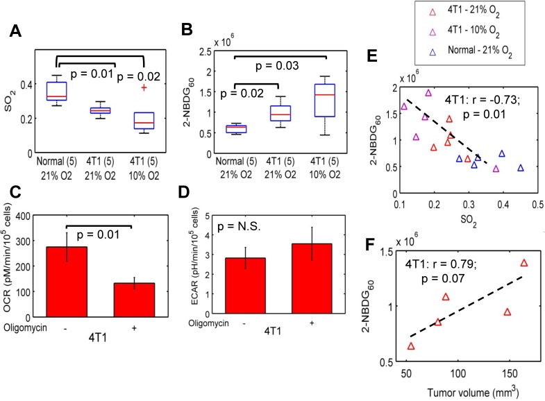
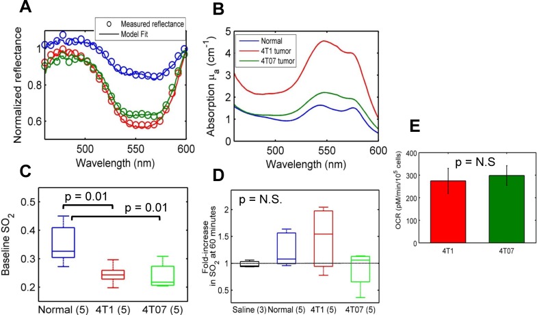
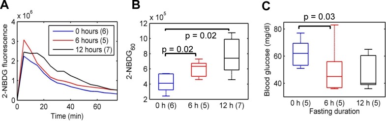
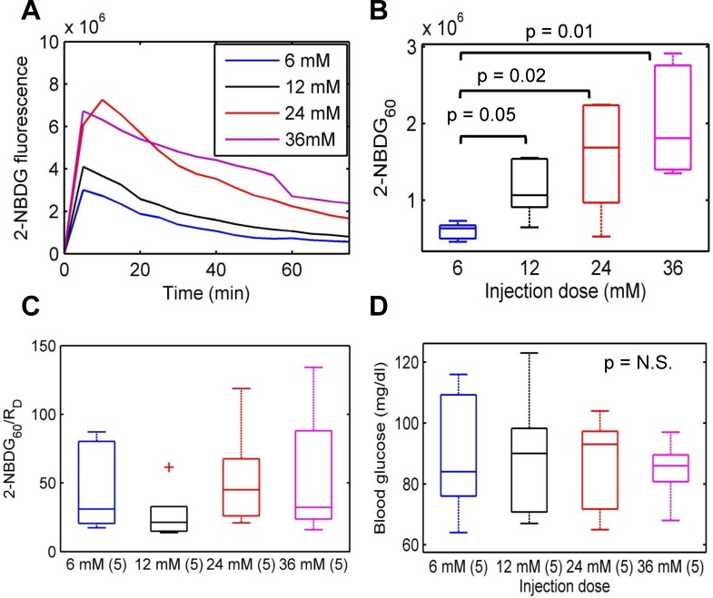
We report the development of non-invasive, fiber-based diffuse optical spectroscopy for simultaneously quantifying vascular oxygenation (SO2) and glucose uptake in solid tumors in vivo. Glucose uptake was measured using a fluorescent glucose analog, 2-[N-(7-nitrobenz-2-oxa-1,3-diaxol-4-yl)amino]-2-deoxyglucose (2-NBDG). Quantification of label-free SO2 and 2-NBDG-fluorescence-based glucose uptake 60 minutes after administration of the tracer (2-NBDG60) was performed using computational models of light-tissue interaction. This study was carried out on normal tissue and 4T1 and 4T07 murine mammary tumor xenografts in vivo. Injection of 2-NBDG did not cause a significant change in optical measurements of SO2, demonstrating its suitability as a functional reporter of tumor glucose uptake. Correction of measured 2-NBDG-fluorescence for the effects of absorption and scattering significantly improved contrast between tumor and normal tissue. The 4T1 and 4T07 tumors showed significantly decreased SO2, and 4T1 tumors demonstrated increased 2-NBDG60 compared with normal tissue (60 minutes after the administration of 2-NBDG when perfusion-mediated effects have cleared). 2-NBDG-fluorescence was found to be highly sensitive to food deprivation-induced reduction in blood glucose levels, demonstrating that this endpoint is indeed sensitive to glycolytic demand. 2-NBDG60 was also found to be linearly related to dose, underscoring the importance of calibrating for dose when comparing across animals or experiments. 4T1 tumors demonstrated an inverse relationship between 2-NBDG60 and SO2 that was consistent with the Pasteur effect, particularly when exposed to hypoxic gas breathing. Our results illustrate the potential of optical spectroscopy to provide valuable information about the metabolic status of tumors, with important implications for cancer prognosis.
我们报告了一种基于光纤的非侵入性漫射光学光谱技术的进展,该技术可在体内同时定量测定实体瘤中的血管氧合(SO2)和葡萄糖摄取。使用荧光葡萄糖类似物2-[N-(7-硝基苯并-2-恶唑-1,3-二氮杂环戊烯-4-基)氨基]-2-脱氧葡萄糖(2-NBDG)来测量葡萄糖摄取。在注射示踪剂(2-NBDG60)60分钟后,使用光与组织相互作用的计算模型对无标记的SO2和基于2-NBDG荧光的葡萄糖摄取进行定量。本研究在正常组织以及4T1和4T07小鼠乳腺肿瘤异种移植模型体内进行。注射2-NBDG并未导致SO2的光学测量值发生显著变化,这表明其适合作为肿瘤葡萄糖摄取的功能性报告分子。针对吸收和散射效应校正测量的2-NBDG荧光,可显著提高肿瘤与正常组织之间的对比度。与正常组织相比(在注射2-NBDG 60分钟后,灌注介导的效应已消除),4T1和4T07肿瘤显示SO2显著降低,4T1肿瘤显示2-NBDG60增加。发现2-NBDG荧光对食物剥夺引起的血糖水平降低高度敏感,表明该终点确实对糖酵解需求敏感。还发现2-NBDG60与剂量呈线性关系,强调了在跨动物或实验进行比较时校准剂量的重要性。4T1肿瘤显示2-NBDG60与SO2之间存在反比关系,这与巴斯德效应一致,尤其是在暴露于低氧气体呼吸时。我们的结果说明了光学光谱技术在提供有关肿瘤代谢状态的有价值信息方面的潜力,这对癌症预后具有重要意义。