Tsou Shang-Hsun, Chen Tzer-Ming, Hsiao Hui-Ting, Chen Yen-Hui
Graduate Institute of Pharmaceutical Sciences, School of Pharmacy, College of Medicine, National Taiwan University, Taipei, Taiwan.
Department of Obstetrics and Gynecology, College of Medicine, National Taiwan University, Taipei, Taiwan.
PLoS One. 2015 Jan 30;10(1):e0116747. doi: 10.1371/journal.pone.0116747. eCollection 2015.
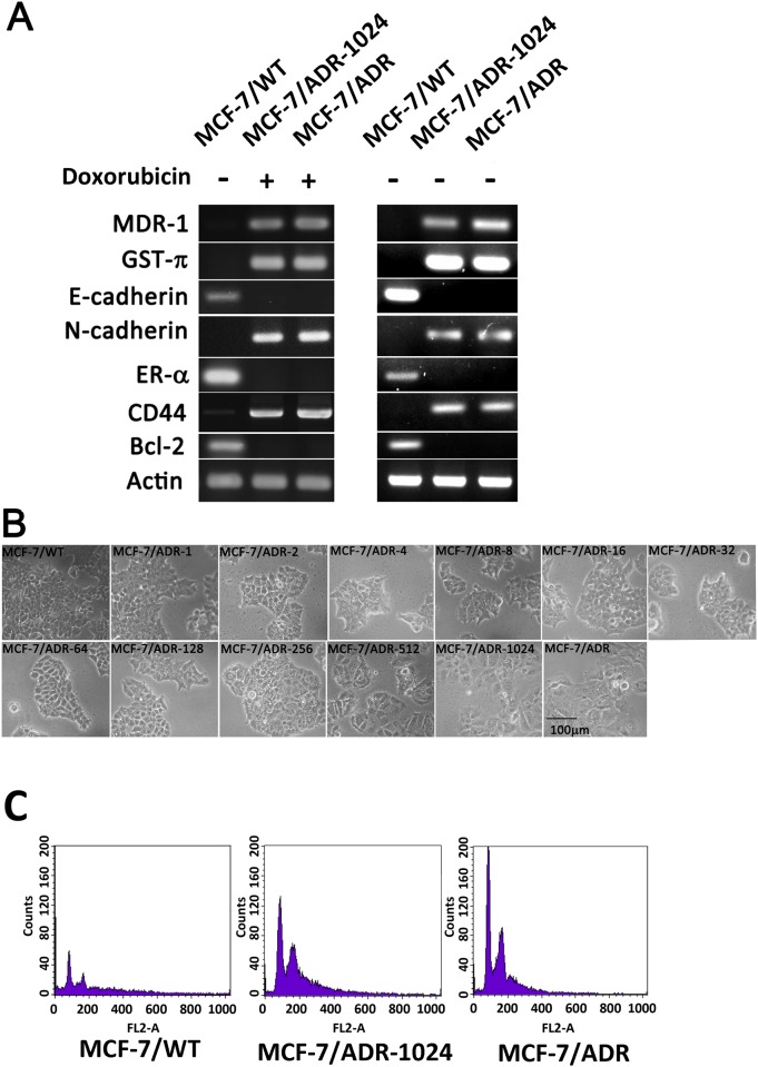
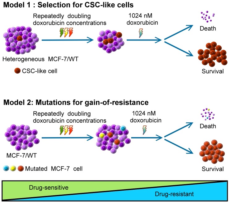
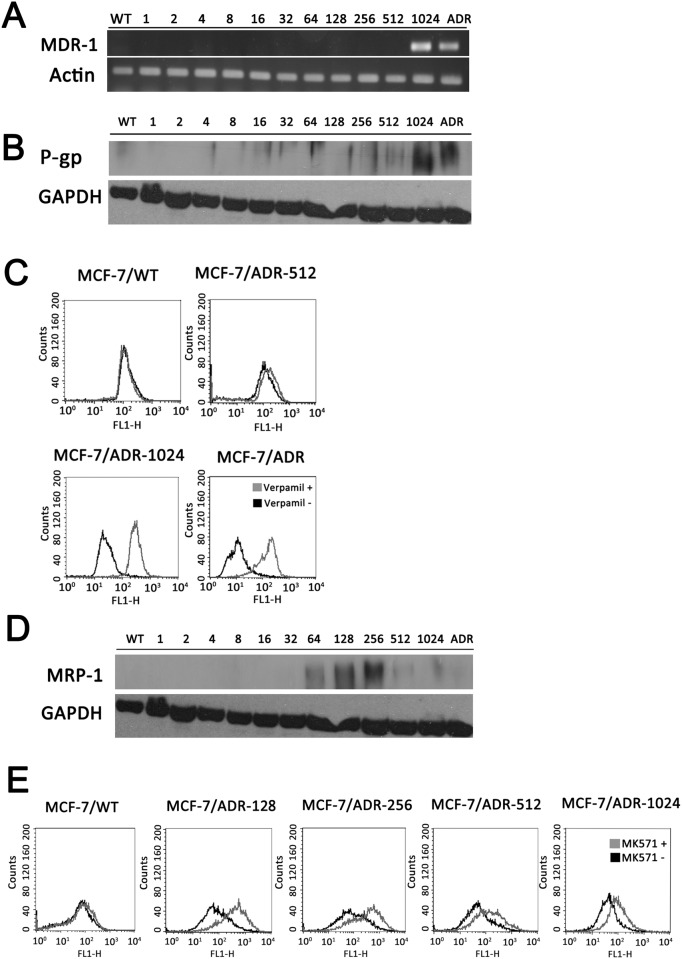
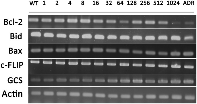
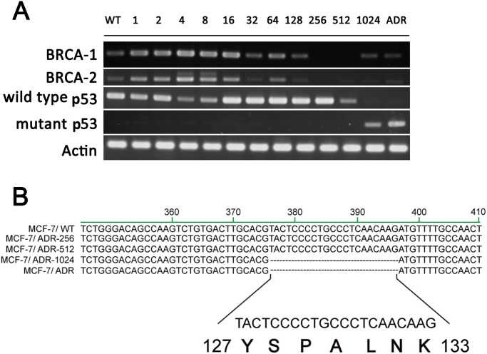
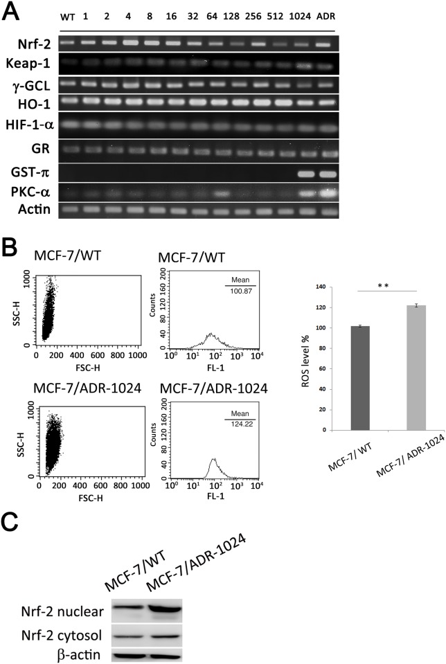
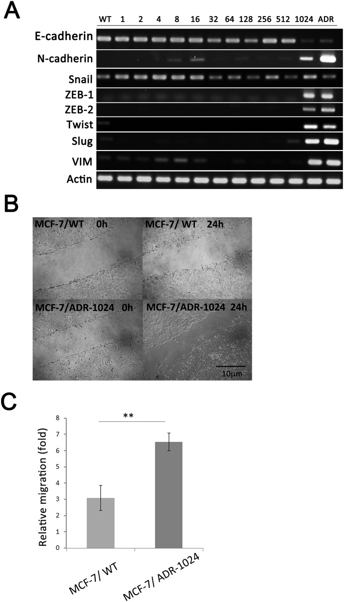
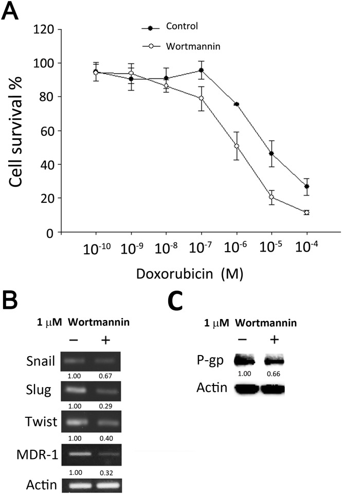
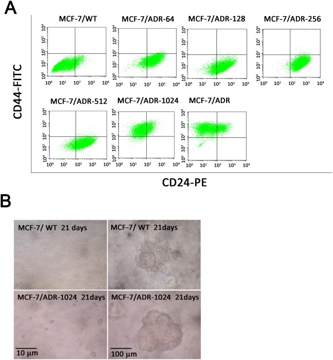
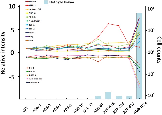
Cellular mechanisms of multidrug resistance (MDR) are related to ABC transporters, apoptosis, antioxidation, drug metabolism, DNA repair and cell proliferation. It remains unclear whether the process of resistance development is programmable. We aimed to study gene expression profiling circumstances in MCF-7 during MDR development. Eleven MCF-7 sublines with incremental doxorubicin resistance were established as a valued tool to study resistance progression. MDR marker P-gp was overexpressed only in cells termed MCF-7/ADR-1024 under the selection dose approaching 1024 nM. MCF-7/ADR-1024 and authentic MCF-7/ADR shared common features in cell morphology and DNA ploidy status. MCF-7/ADR-1024 and authentic MCF-7/ADR down regulated repair genes BRCA1/2 and wild type p53, apoptosis-related gene Bcl-2 and epithelial-mesenchymal transition (EMT) epithelial marker gene E-cadherin. While detoxifying enzymes glutathione-S transferase-π and protein kinase C-α were up-regulated. The genes involving in EMT mesenchymal formation were also overexpressed, including N-cadherin, vimentin and the E-cadherin transcription reppressors Slug, Twist and ZEB1/2. PI3K/AKT inhibitor wortmannin suppressed expression of Slug, Twist and mdr1. Mutant p53 with a deletion at codons 127-133 markedly appeared in MCF-7/ADR-1024 and authentic MCF-7/ADR as well. In addition, MCF-7/ADR-1024 cells exerted CSC-like cell surface marker CD44 high/CD24 low and form mammospheres. Overall, results suggest that resistance marker P-gp arises owing to turn on/off or mutation of the genes involved in DNA repair, apoptosis, detoxifying enzymes, EMT and ABC transporters at a turning point (1.024 μM doxorubicin challenge). Behind this point, no obvious alterations were found in most tested genes. Selection for CSC-like cells under this dose may importantly attribute to propagation of the population presenting invasive properties and drug resistance. We thereby suggest two models in the induction of drug resistance. Model 1: Selection for CSC-like cells. Model 2: Mutations for gain-of resistance. Either model 1 or model 2 requires doxorubicin dose approaching 1 μM to alter gene regulation.
多药耐药(MDR)的细胞机制与ABC转运蛋白、细胞凋亡、抗氧化、药物代谢、DNA修复及细胞增殖有关。耐药性发展过程是否具有可程序性仍不清楚。我们旨在研究MCF-7细胞在MDR发展过程中的基因表达谱情况。建立了11个对阿霉素耐药性递增的MCF-7亚系,作为研究耐药性进展的重要工具。在接近1024 nM的选择剂量下,MDR标志物P-糖蛋白仅在称为MCF-7/ADR-1024的细胞中过表达。MCF-7/ADR-1024与正宗的MCF-7/ADR在细胞形态和DNA倍体状态上具有共同特征。MCF-7/ADR-1024和正宗的MCF-7/ADR下调了修复基因BRCA1/2和野生型p53、凋亡相关基因Bcl-2以及上皮-间质转化(EMT)上皮标志物基因E-钙黏蛋白。同时,解毒酶谷胱甘肽-S转移酶-π和蛋白激酶C-α上调。参与EMT间质形成的基因也过表达,包括N-钙黏蛋白、波形蛋白以及E-钙黏蛋白转录抑制因子Slug、Twist和ZEB1/2。PI3K/AKT抑制剂渥曼青霉素抑制了Slug、Twist和mdr1的表达。在MCF-7/ADR-1024和正宗的MCF-7/ADR中也明显出现了密码子127 - 133缺失的突变型p53。此外,MCF-7/ADR-1024细胞呈现CSC样细胞表面标志物CD44高/CD24低,并形成乳腺球。总体而言,结果表明耐药标志物P-糖蛋白的出现是由于在一个转折点(1.024 μM阿霉素刺激)时,参与DNA修复、细胞凋亡、解毒酶、EMT和ABC转运蛋白的基因开启/关闭或发生突变。在此转折点之后,大多数检测基因未发现明显变化。在此剂量下对CSC样细胞的选择可能对具有侵袭性和耐药性的细胞群体的增殖有重要影响。因此,我们提出了两种耐药诱导模型。模型1:对CSC样细胞的选择。模型2:获得性耐药突变。模型1或模型2都需要阿霉素剂量接近1 μM来改变基因调控。