Lo Yi-Wen, Lin Szu-Ting, Chang Shing-Jyh, Chan Chia-Hao, Lyu Kevin W, Chang Jo-Fan, May Eugenie Wong Soon, Lin Dai-Ying, Chou Hsiu-Chuan, Chan Hong-Lin
Department of Applied Science, National Hsinchu University of Education, Hsinchu, Taiwan.
J Cell Mol Med. 2015 Apr;19(4):744-59. doi: 10.1111/jcmm.12388. Epub 2015 Jan 30.
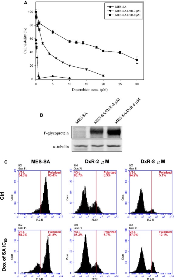
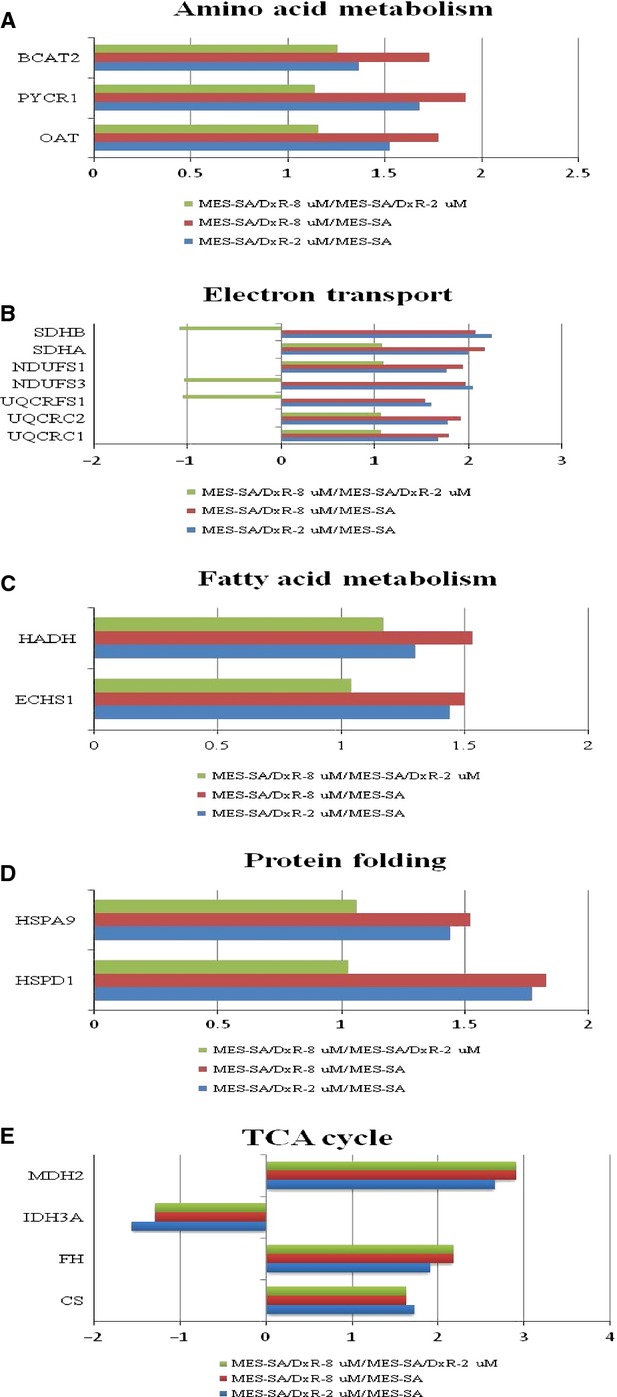
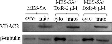
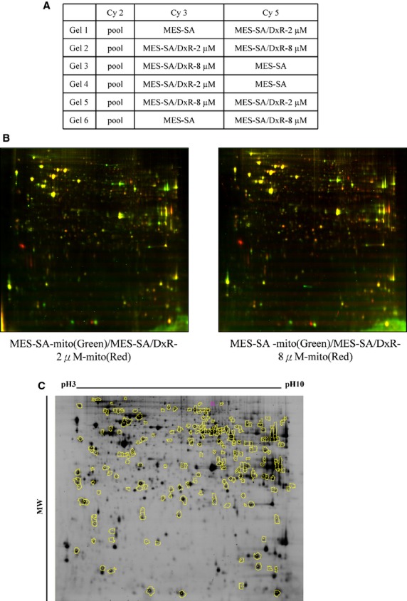
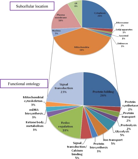
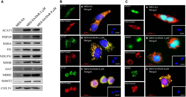
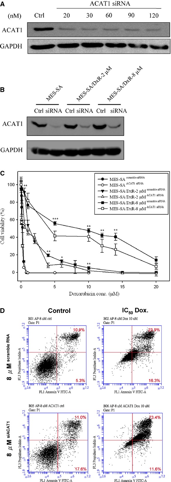
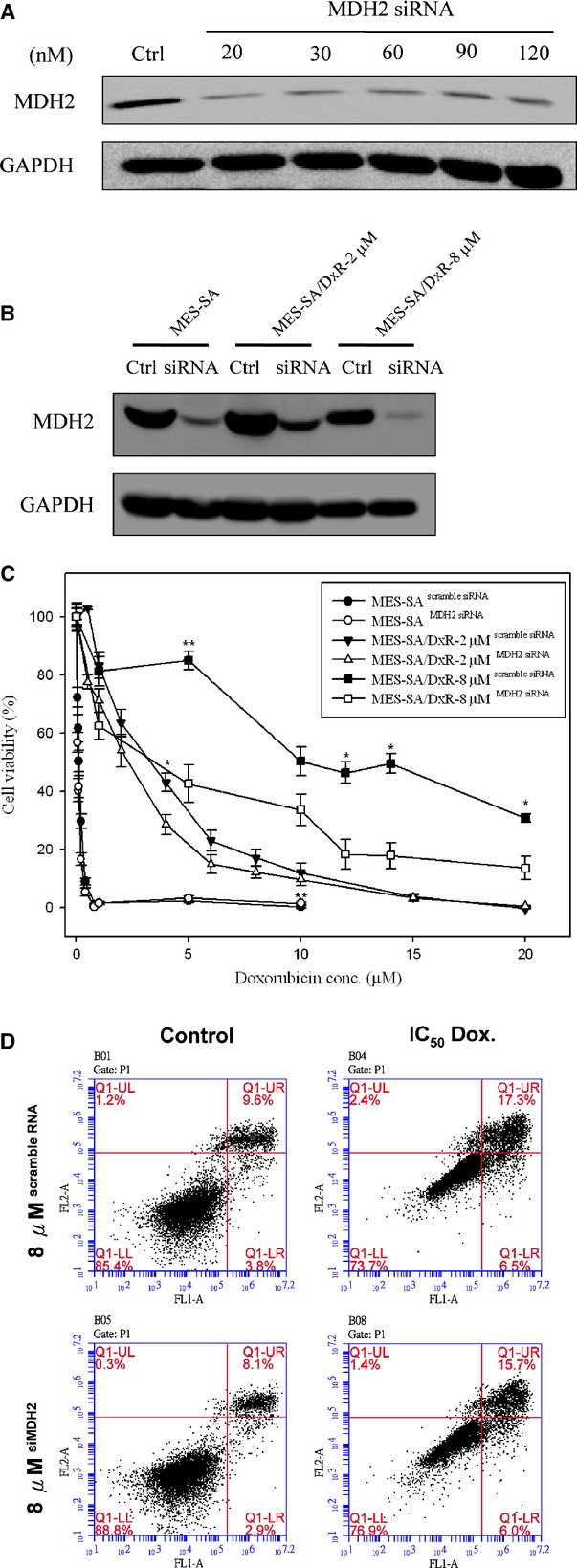
Mitochondria are key organelles in mammary cells in responsible for a number of cellular functions including cell survival and energy metabolism. Moreover, mitochondria are one of the major targets under doxorubicin treatment. In this study, low-abundant mitochondrial proteins were enriched for proteomic analysis with the state-of-the-art two-dimensional differential gel electrophoresis (2D-DIGE) and matrix-assistant laser desorption ionization time-of-flight mass spectrometry (MALDI-TOF MS) strategy to compare and identify the mitochondrial protein profiling changes in response to the development of doxorubicin resistance in human uterine cancer cells. The mitochondrial proteomic results demonstrate more than fifteen hundred protein features were resolved from the equal amount pooled of three purified mitochondrial proteins and 101 differentially expressed spots were identified. In which, 39 out of these 101 identified proteins belong to mitochondrial proteins. Mitochondrial proteins such as acetyl-CoA acetyltransferase (ACAT1) and malate dehydrogenase (MDH2) have not been reported with the roles on the formation of doxorubicin resistance in our knowledge. Further studies have used RNA interference and cell viability analysis to evidence the essential roles of ACAT1 and MDH2 on their potency in the formation of doxorubicin resistance through increased cell viability and decreased cell apoptosis during doxorubicin treatment. To sum up, our current mitochondrial proteomic approaches allowed us to identify numerous proteins, including ACAT1 and MDH2, involved in various drug-resistance-forming mechanisms. Our results provide potential diagnostic markers and therapeutic candidates for the treatment of doxorubicin-resistant uterine cancer.
线粒体是乳腺细胞中的关键细胞器,负责多种细胞功能,包括细胞存活和能量代谢。此外,线粒体是阿霉素治疗的主要靶点之一。在本研究中,采用先进的二维差异凝胶电泳(2D-DIGE)和基质辅助激光解吸电离飞行时间质谱(MALDI-TOF MS)策略对低丰度线粒体蛋白进行富集,以进行蛋白质组学分析,比较和鉴定人子宫癌细胞对阿霉素耐药性发展过程中线粒体蛋白质谱的变化。线粒体蛋白质组学结果表明,从三种纯化的线粒体蛋白等量混合样本中解析出了1500多个蛋白质特征,并鉴定出101个差异表达斑点。其中,这101个已鉴定蛋白质中有39个属于线粒体蛋白。据我们所知,乙酰辅酶A乙酰转移酶(ACAT1)和苹果酸脱氢酶(MDH2)等线粒体蛋白在阿霉素耐药形成中的作用尚未见报道。进一步的研究使用RNA干扰和细胞活力分析,以证明ACAT1和MDH2在阿霉素治疗期间通过提高细胞活力和降低细胞凋亡来形成阿霉素耐药性的过程中发挥的重要作用。综上所述,我们目前的线粒体蛋白质组学方法使我们能够鉴定出许多参与各种耐药形成机制的蛋白质,包括ACAT1和MDH2。我们的结果为阿霉素耐药性子宫癌的治疗提供了潜在的诊断标志物和治疗候选物。