Castorina Alessandro, Waschek James A, Marzagalli Rubina, Cardile Venera, Drago Filippo
Department of Biomedical Sciences and Biotechnologies, Section of Human Anatomy and Histology, University of Catania, Catania, Italy.
Semel Institute/Department of Psychiatry, David Geffen School of Medicine, University of California Los Angeles, Los Angeles, California, United States of America.
PLoS One. 2015 Feb 6;10(2):e0117799. doi: 10.1371/journal.pone.0117799. eCollection 2015.
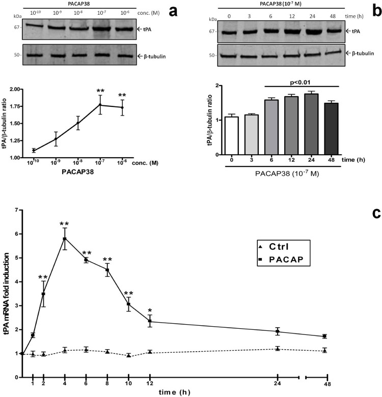
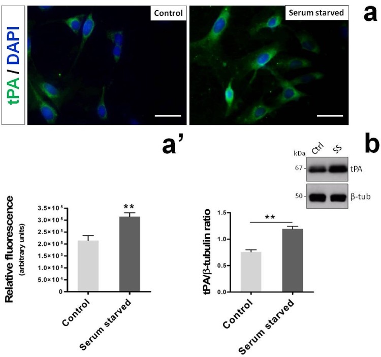
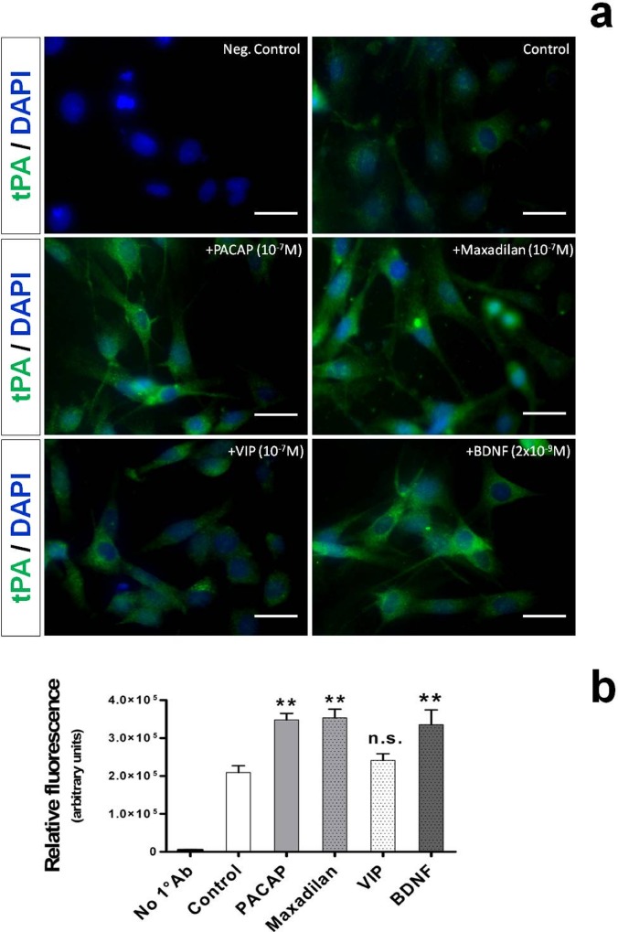
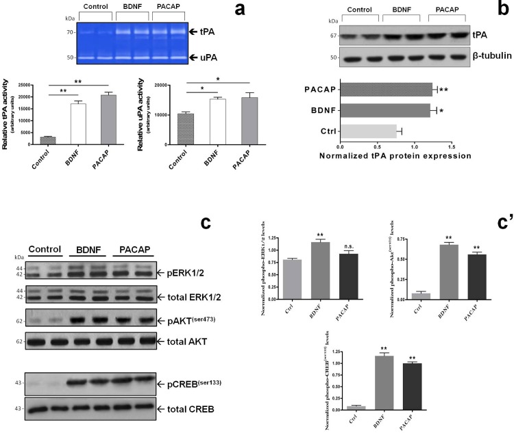
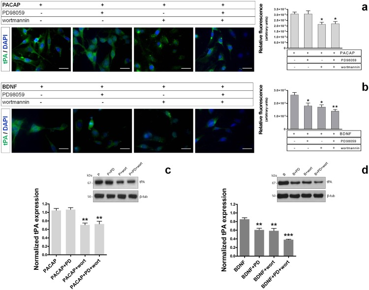
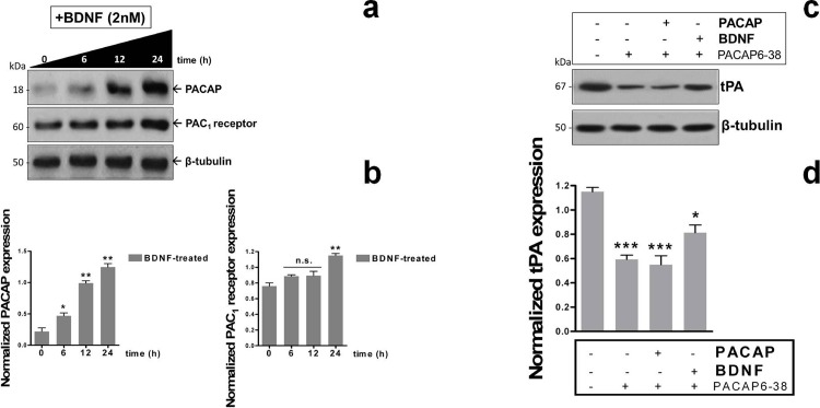
Regeneration of peripheral nerves depends on the abilities of rejuvenating axons to migrate at the injury site through cellular debris and altered extracellular matrix, and then grow along the residual distal nerve sheath conduit and reinnervate synaptic targets. Considerable evidence suggest that glial cells participate in this process, although the mechanisms remain to be clarified. In cell culture, regenerating neurites secrete PACAP, a peptide shown to induce the expression of the protease tissue plasminogen activator (tPA) in neural cell types. In the present studies, we tested the hypothesis that PACAP can stimulate peripheral glial cells to produce tPA. More specifically, we addressed whether or not PACAP promoted the expression and activity of tPA in the Schwann cell line RT4-D6P2T, which shares biochemical and physical properties with Schwann cells. We found that PACAP dose- and time-dependently stimulated tPA expression both at the mRNA and protein level. Such effect was mimicked by maxadilan, a potent PAC1 receptor agonist, but not by the PACAP-related homolog VIP, suggesting a PAC1-mediated function. These actions appeared to be mediated at least in part by the Akt/CREB signaling cascade because wortmannin, a PI3K inhibitor, prevented peptide-driven CREB phosphorylation and tPA increase. Interestingly, treatment with BDNF mimicked PACAP actions on tPA, but acted through both the Akt and MAPK signaling pathways, while causing a robust increase in PACAP and PAC1 expression. PACAP6-38 totally blocked PACAP-driven tPA expression and in part hampered BDNF-mediated effects. We conclude that PACAP, acting through PAC1 receptors, stimulates tPA expression and activity in a Akt/CREB-dependent manner to promote proteolytic activity in Schwann-cell like cultures.
周围神经的再生取决于轴突在损伤部位通过细胞碎片和改变的细胞外基质进行迁移的能力,然后沿着残留的远端神经鞘管生长并重新支配突触靶点。大量证据表明神经胶质细胞参与了这一过程,尽管其机制仍有待阐明。在细胞培养中,再生的神经突分泌垂体腺苷酸环化酶激活肽(PACAP),这是一种已被证明能诱导神经细胞类型中蛋白酶组织型纤溶酶原激活剂(tPA)表达的肽。在本研究中,我们检验了PACAP能刺激周围神经胶质细胞产生tPA这一假说。更具体地说,我们探讨了PACAP是否促进了雪旺氏细胞系RT4-D6P2T中tPA的表达和活性,该细胞系与雪旺氏细胞具有相同的生化和物理特性。我们发现PACAP在mRNA和蛋白质水平上均呈剂量和时间依赖性地刺激tPA表达。强效PAC1受体激动剂马西替坦可模拟这种效应,但PACAP相关同系物血管活性肠肽(VIP)则不能,这表明是由PAC1介导的功能。这些作用似乎至少部分是由Akt/CREB信号级联介导的,因为磷脂酰肌醇-3激酶(PI3K)抑制剂渥曼青霉素可阻止肽驱动的CREB磷酸化和tPA增加。有趣的是,脑源性神经营养因子(BDNF)处理可模拟PACAP对tPA的作用,但通过Akt和丝裂原活化蛋白激酶(MAPK)信号通路发挥作用,同时导致PACAP和PAC1表达显著增加。PACAP6-38完全阻断了PACAP驱动的tPA表达,并部分阻碍了BDNF介导的效应。我们得出结论,PACAP通过PAC1受体发挥作用,以Akt/CREB依赖性方式刺激tPA表达和活性,从而促进雪旺氏细胞样培养物中的蛋白水解活性。