Severgnini Mara, de Denaro Mario, Bortul Marina, Vidali Cristiana, Beorchia Aulo
Department of Medical Physics, A.O.U. "Ospedali Riuniti" di Trieste, Italy.
J Appl Clin Med Phys. 2014 Jan 8;16(1):5065. doi: 10.1120/jacmp.v16i1.5065.
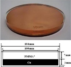
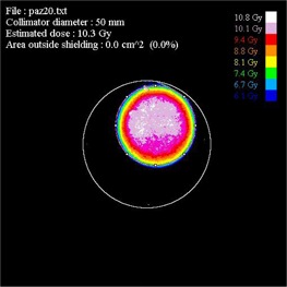
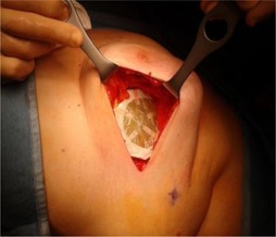
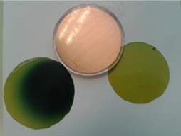
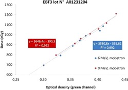
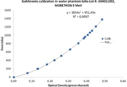
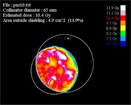
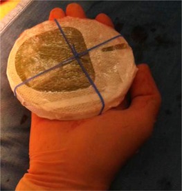
Intraoperative electron radiation therapy (IOERT) cannot usually benefit, as conventional external radiotherapy, from software systems of treatment planning based on computed tomography and from common dose verify procedures. For this reason, in vivo film dosimetry (IVFD) proves to be an effective methodology to evaluate the actual radiation dose delivered to the target. A practical method for IVFD during breast IOERT was carried out to improve information on the dose actually delivered to the tumor target and on the alignment of the shielding disk with respect to the electron beam. Two EBT3 GAFCHROMIC films have been positioned on the two sides of the shielding disk in order to obtain the dose maps at the target and beyond the disk. Moreover the postprocessing analysis of the dose distribution measured on the films provides a quantitative estimate of the misalignment between the collimator and the disk. EBT3 radiochromic films have been demonstrated to be suitable dosimeters for IVD due to their linear dose-optical density response in a narrow range around the prescribed dose, as well as their capability to be fixed to the shielding disk without giving any distortion in the dose distribution. Off-line analysis of the radiochromic film allowed absolute dose measurements and this is indeed a very important verification of the correct exposure to the target organ, as well as an estimate of the dose to the healthy tissue underlying the shielding. These dose maps allow surgeons and radiation oncologists to take advantage of qualitative and quantitative feedback for setting more accurate treatment strategies and further optimized procedures. The proper alignment using elastic bands has improved the absolute dose accuracy and the collimator disk alignment by more than 50%.
术中电子放射治疗(IOERT)通常无法像传统外照射放疗那样,受益于基于计算机断层扫描的治疗计划软件系统和常规剂量验证程序。因此,体内胶片剂量测定法(IVFD)被证明是一种评估实际输送到靶区的辐射剂量的有效方法。开展了一种用于乳腺IOERT期间IVFD的实用方法,以改善有关实际输送到肿瘤靶区的剂量以及屏蔽盘相对于电子束的对准情况的信息。已将两张EBT3 GAFCHROMIC胶片放置在屏蔽盘的两侧,以便获取靶区和屏蔽盘之外的剂量分布图。此外,对胶片上测量的剂量分布进行的后处理分析提供了准直器与屏蔽盘之间未对准情况的定量估计。EBT3放射变色胶片已被证明是适用于IVD的剂量计,这是由于其在规定剂量附近的窄范围内具有线性剂量-光密度响应,以及其能够固定在屏蔽盘上而不会在剂量分布中产生任何畸变。对放射变色胶片的离线分析允许进行绝对剂量测量,这确实是对靶器官正确照射的非常重要的验证,也是对屏蔽下方健康组织剂量的估计。这些剂量分布图使外科医生和放射肿瘤学家能够利用定性和定量反馈来制定更准确的治疗策略和进一步优化的程序。使用弹性带进行适当对准使绝对剂量精度和准直器盘对准提高了50%以上。