Del Rio Carlos L, Clymer Bradley D, Billman George E
Department of Physiology and Cell Biology, The Ohio State University Columbus, OH, USA ; Department of Electrical and Computer Engineering, The Ohio State University Columbus, OH, USA ; Safety Pharmacology, QTest Labs Columbus, OH, USA.
Department of Electrical and Computer Engineering, The Ohio State University Columbus, OH, USA ; Biomedical Engineering, The Ohio State University Columbus, OH, USA.
Front Physiol. 2015 Feb 5;6:25. doi: 10.3389/fphys.2015.00025. eCollection 2015.
Autonomic neural activation during cardiac stress testing is an established risk-stratification tool in post-myocardial infarction (MI) patients. However, autonomic activation can also modulate myocardial electrotonic coupling, a known factor to contribute to the genesis of arrhythmias. The present study tested the hypothesis that exercise-induced autonomic neural activation modulates electrotonic coupling (as measured by myocardial electrical impedance, MEI) in post-MI animals shown to be susceptible or resistant to ventricular fibrillation (VF).
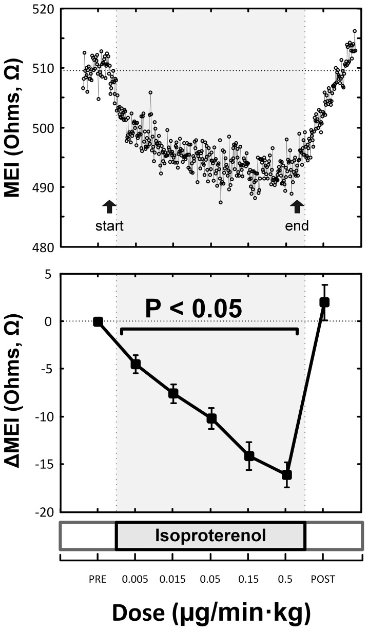
Dogs (n = 25) with healed MI instrumented for MEI measurements were trained to run on a treadmill and classified based on their susceptibility to VF (12 susceptible, 9 resistant). MEI and ECGs were recorded during 6-stage exercise tests (18 min/test; peak: 6.4 km/h @ 16%) performed under control conditions, and following complete β-adrenoceptor (β-AR) blockade (propranolol); MEI was also measured at rest during escalating β-AR stimulation (isoproterenol) or overdrive-pacing.
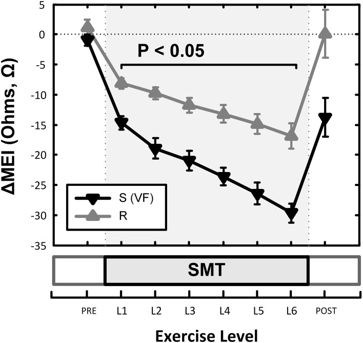
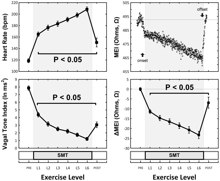
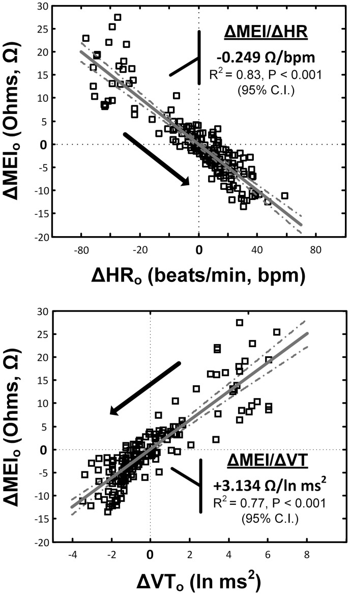
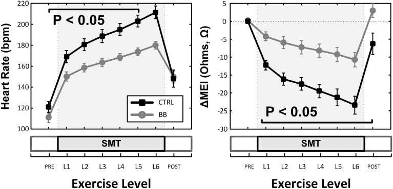
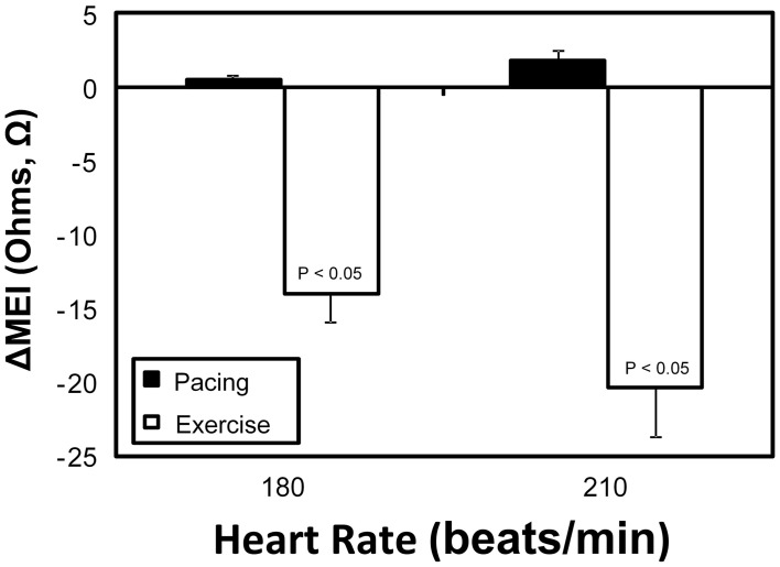
Exercise progressively increased heart rate (HR) and reduced heart rate variability (HRV). In parallel, MEI decreased gradually (enhanced electrotonic coupling) with exercise; at peak exercise, MEI was reduced by 5.3 ± 0.4% (or -23 ± 1.8Ω, P < 0.001). Notably, exercise-mediated electrotonic changes were linearly predicted by the degree of autonomic activation, as indicated by changes in either HR or in HRV (P < 0.001). Indeed, β-AR blockade attenuated the MEI response to exercise while direct β-AR stimulation (at rest) triggered MEI decreases comparable to those observed during exercise; ventricular pacing had no significant effects on MEI. Finally, animals prone to VF had a significantly larger MEI response to exercise.
These data suggest that β-AR activation during exercise can acutely enhance electrotonic coupling in the myocardium, particularly in dogs susceptible to ischemia-induced VF.
心脏应激测试期间的自主神经激活是心肌梗死(MI)后患者既定的风险分层工具。然而,自主神经激活也可调节心肌电紧张耦联,这是导致心律失常发生的一个已知因素。本研究检验了以下假设:运动诱导的自主神经激活可调节MI后动物的电紧张耦联(通过心肌电阻抗,MEI测量),这些动物表现出对室颤(VF)敏感或耐受。
对25只已愈合MI且植入用于MEI测量装置的犬进行训练,使其在跑步机上跑步,并根据它们对VF的易感性进行分类(12只敏感,9只耐受)。在对照条件下以及完全β-肾上腺素能受体(β-AR)阻断(普萘洛尔)后,进行6阶段运动测试(18分钟/测试;峰值:6.4公里/小时@16%)期间记录MEI和心电图;在递增β-AR刺激(异丙肾上腺素)或超速起搏期间的静息状态下也测量MEI。
运动使心率(HR)逐渐增加,心率变异性(HRV)降低。同时,随着运动MEI逐渐降低(电紧张耦联增强);在运动峰值时,MEI降低了5.3±0.4%(或-23±1.8Ω,P<0.001)。值得注意的是,运动介导的电紧张变化可由自主神经激活程度线性预测,这通过HR或HRV的变化表明(P<0.001)。实际上,β-AR阻断减弱了MEI对运动的反应,而直接β-AR刺激(静息时)引发的MEI降低与运动期间观察到的相当;心室起搏对MEI无显著影响。最后,易发生VF的动物对运动的MEI反应明显更大。
这些数据表明运动期间β-AR激活可急性增强心肌中的电紧张耦联,特别是在易发生缺血诱导VF的犬中。