Szeiffova Bacova Barbara, Viczenczova Csilla, Andelova Katarina, Sykora Matus, Chaudagar Kiranj, Barancik Miroslav, Adamcova Michaela, Knezl Vladimir, Egan Benova Tamara, Weismann Peter, Slezak Jan, Tribulova Narcisa
Centre of Experimental Medicine, SAS, 84104 Bratislava, Slovakia.
Research Center for Molecular Medicine of the Austrian Academy of Sciences, A-1090 Vienna, Austria.
Antioxidants (Basel). 2020 Jun 22;9(6):546. doi: 10.3390/antiox9060546.
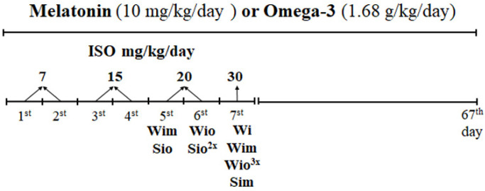
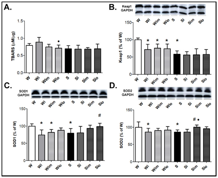
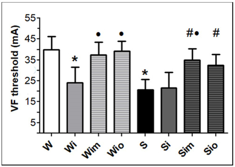
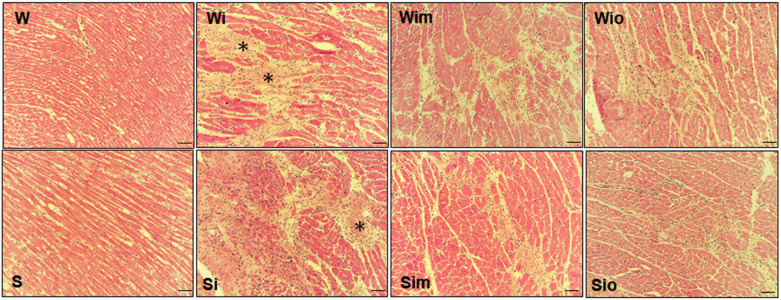
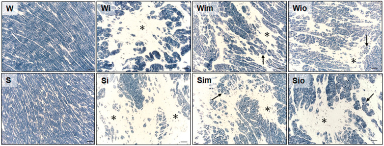
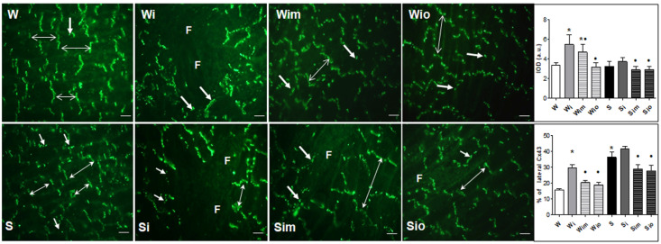
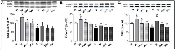
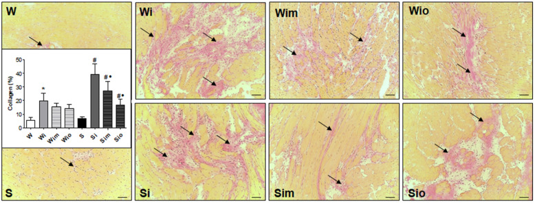
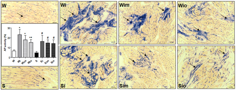
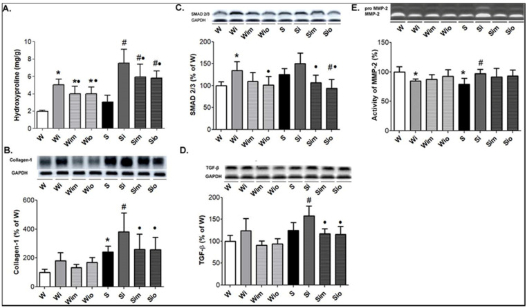
Cardiac β-adrenergic overstimulation results in oxidative stress, hypertrophy, ischemia, lesion, and fibrosis rendering the heart vulnerable to malignant arrhythmias. We aimed to explore the anti-arrhythmic efficacy of the anti-oxidative and anti-inflammatory compounds, melatonin, and omega-3, and their mechanisms of actions in normotensive and hypertensive rats exposed to isoproterenol (ISO) induced β-adrenergic overdrive. Eight-month-old, male SHR, and Wistar rats were injected during 7 days with ISO (cumulative dose, 118 mg/kg). ISO rats were either untreated or concomitantly treated with melatonin (10 mg/kg/day) or omega-3 (Omacor, 1.68 g/kg/day) until 60 days of ISO withdrawal and compared to non-ISO controls. Findings showed that both melatonin and omega-3 increased threshold current to induce ventricular fibrillation (VF) in ISO rats regardless of the strain. Prolonged treatment with these compounds resulted in significant suppression of ISO-induced extracellular matrix alterations, as indicated by reduced areas of diffuse fibrosis and decline of hydroxyproline, collagen-1, SMAD2/3, and TGF-β1 protein levels. Importantly, the highly pro-arrhythmic ISO-induced disordered cardiomyocyte distribution of electrical coupling protein, connexin-43 (Cx43), and its remodeling (lateralization) were significantly attenuated by melatonin and omega-3 in Wistar as well as SHR hearts. In parallel, both compounds prevented the post-ISO-related increase in Cx43 variant phosphorylated at serine 368 along with PKCε, which are known to modulate Cx43 remodeling. Melatonin and omega-3 increased SOD1 or SOD2 protein levels in ISO-exposed rats of both strains. Altogether, the results indicate that anti-arrhythmic effects of melatonin and omega-3 might be attributed to the protection of myocardial Cx43 topology and suppression of fibrosis in the setting of oxidative stress induced by catecholamine overdrive in normotensive and hypertensive rats.
心脏β-肾上腺素能过度刺激会导致氧化应激、肥大、缺血、损伤和纤维化,使心脏易患恶性心律失常。我们旨在探讨抗氧化和抗炎化合物褪黑素和ω-3在暴露于异丙肾上腺素(ISO)诱导的β-肾上腺素能过度驱动的正常血压和高血压大鼠中的抗心律失常疗效及其作用机制。8月龄雄性自发性高血压大鼠(SHR)和Wistar大鼠连续7天注射ISO(累积剂量118 mg/kg)。ISO大鼠要么不治疗,要么同时用褪黑素(10 mg/kg/天)或ω-3(Omacor,1.68 g/kg/天)治疗,直至停止注射ISO 60天,并与未注射ISO的对照组进行比较。研究结果表明,无论何种品系,褪黑素和ω-3均可提高ISO大鼠诱发心室颤动(VF)的阈电流。用这些化合物进行长期治疗可显著抑制ISO诱导的细胞外基质改变,表现为弥漫性纤维化面积减少以及羟脯氨酸、胶原蛋白-1、SMAD2/3和TGF-β1蛋白水平下降。重要的是,在Wistar大鼠和SHR大鼠的心脏中,褪黑素和ω-3可显著减轻ISO诱导的促心律失常的电偶联蛋白连接蛋白43(Cx43)的无序心肌细胞分布及其重塑(侧向化)。同时,这两种化合物均可防止ISO后与丝氨酸368磷酸化的Cx43变体以及蛋白激酶Cε(PKCε)相关的增加,已知它们可调节Cx43重塑。褪黑素和ω-3可提高两种品系暴露于ISO的大鼠中SOD1或SOD2蛋白水平。总之,结果表明,褪黑素和ω-3的抗心律失常作用可能归因于在正常血压和高血压大鼠中儿茶酚胺过度驱动诱导的氧化应激情况下对心肌Cx43拓扑结构的保护和纤维化的抑制。