Himel Herman D, Garny Alan, Noble Penelope J, Wadgaonkar Raj, Savarese Joseph, Liu Nian, Bub Gil, El-Sherif Nabil
G. Bub: Department of Physiology Anatomy and Genetics, Sherrington Building Room C-33, University of Oxford, Oxford, Oxfordshire, UK, OX1 3PT.
J Physiol. 2013 Nov 1;591(21):5357-64. doi: 10.1113/jphysiol.2013.262923. Epub 2013 Sep 9.
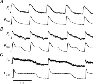
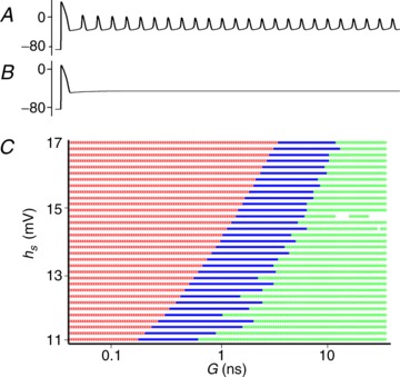
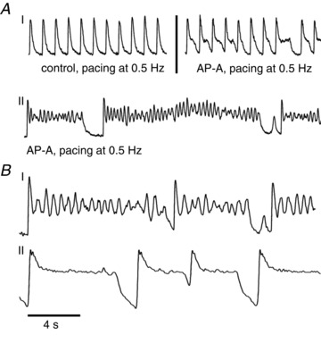
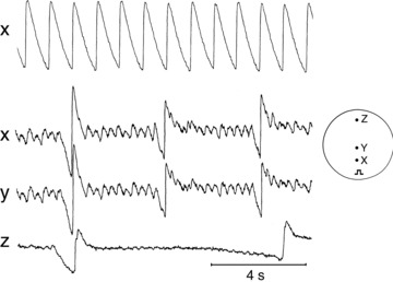
Pathologies that result in early afterdepolarizations (EADs) are a known trigger for tachyarrhythmias, but the conditions that cause surrounding tissue to conduct or suppress EADs are poorly understood. Here we introduce a cell culture model of EAD propagation consisting of monolayers of cultured neonatal rat ventricular myocytes treated with anthopleurin-A (AP-A). AP-A-treated monolayers display a cycle length dependent prolongation of action potential duration (245 ms untreated, vs. 610 ms at 1 Hz and 1200 ms at 0.5 Hz for AP-A-treated monolayers). In contrast, isolated single cells treated with AP-A develop prominent irregular oscillations with a frequency of 2.5 Hz, and a variable prolongation of the action potential duration of up to several seconds. To investigate whether electrotonic interactions between coupled cells modulates EAD formation, cell connectivity was reduced by RNA silencing gap junction Cx43. In contrast to well-connected monolayers, gap junction silenced monolayers display bradycardia-dependent plateau oscillations consistent with EADs. Further, simulations of a cell displaying EADs electrically connected to a cell with normal action potentials show a coupling strength-dependent suppression of EADs consistent with the experimental results. These results suggest that electrotonic effects may play a critical role in EAD-mediated arrhythmogenesis.
导致早期后去极化(EADs)的病理状况是已知的快速性心律失常触发因素,但导致周围组织传导或抑制EADs的条件却知之甚少。在此,我们引入了一种EAD传播的细胞培养模型,该模型由用海葵毒素-A(AP-A)处理的新生大鼠心室肌细胞单层组成。经AP-A处理的单层细胞表现出动作电位时程的周期长度依赖性延长(未处理时为245毫秒,而经AP-A处理的单层细胞在1赫兹时为610毫秒,在0.5赫兹时为1200毫秒)。相比之下,用AP-A处理的分离单细胞会出现频率为2.5赫兹的明显不规则振荡,以及动作电位时程长达数秒的可变延长。为了研究耦合细胞之间的电紧张相互作用是否调节EAD的形成,通过RNA沉默缝隙连接蛋白Cx43来降低细胞连接性。与连接良好的单层细胞相比,缝隙连接沉默的单层细胞表现出与EAD一致的心动过缓依赖性平台振荡。此外,对一个显示EAD的细胞与一个具有正常动作电位的细胞电连接的模拟显示,EAD的抑制与耦合强度有关,这与实验结果一致。这些结果表明,电紧张效应可能在EAD介导的心律失常发生中起关键作用。