Scarff Charlotte A, Almeida Bruno, Fraga Joana, Macedo-Ribeiro Sandra, Radford Sheena E, Ashcroft Alison E
From the ‡Astbury Centre for Structural Molecular Biology, School of Molecular and Cellular Biology, University of Leeds, Leeds, LS2 9JT, UK;
§IBMC-Instituto de Biologia Molecular e Celular, Universidade do Porto, 4150-4180 Porto, Portugal.
Mol Cell Proteomics. 2015 May;14(5):1241-53. doi: 10.1074/mcp.M114.044610. Epub 2015 Feb 19.
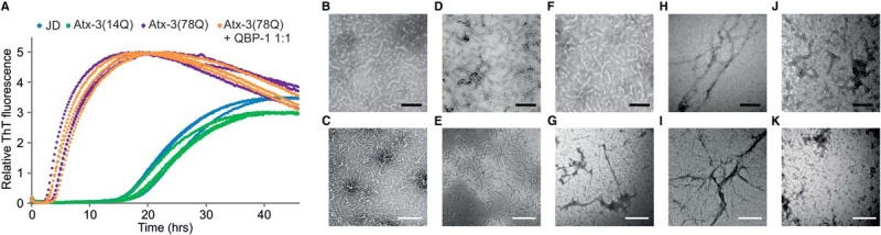
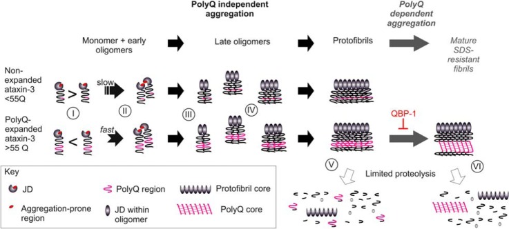
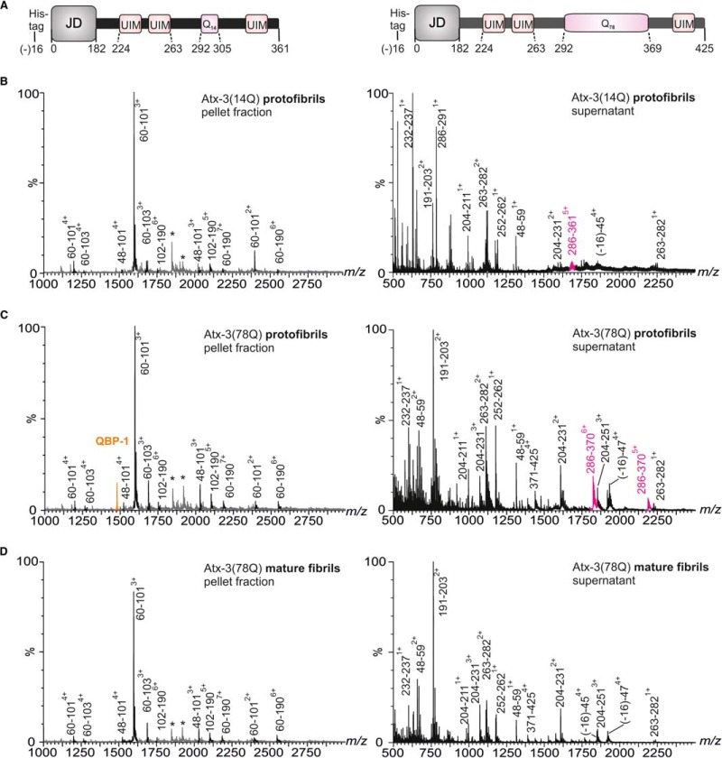
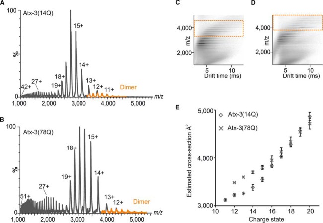
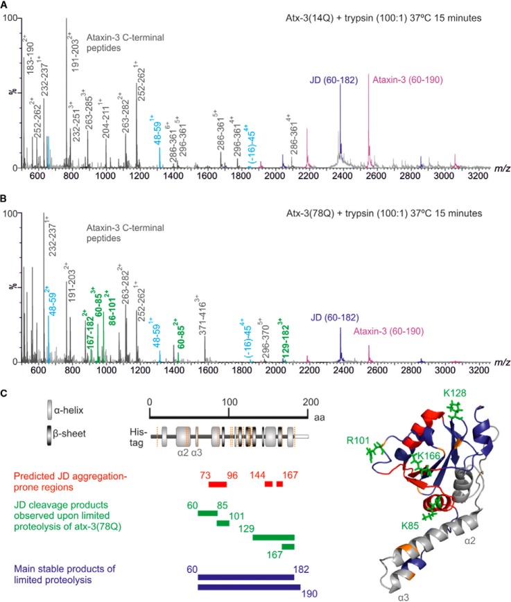
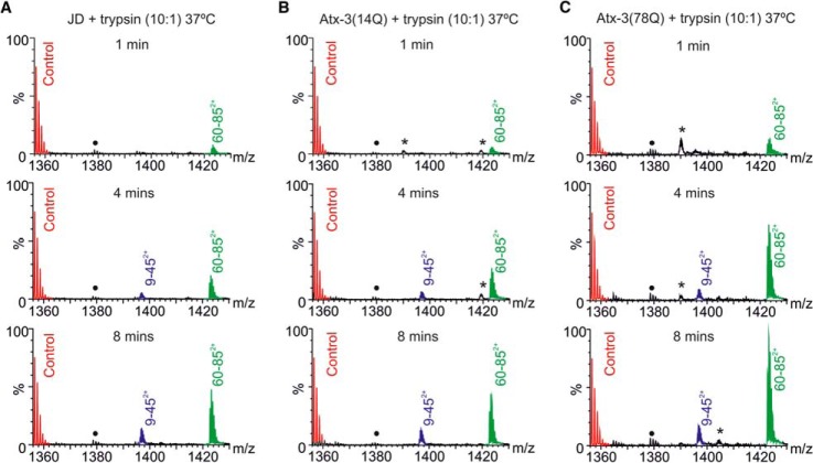
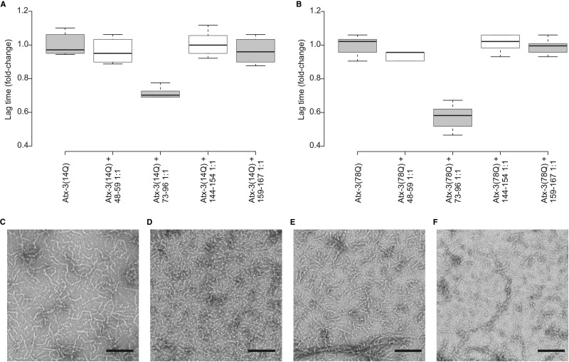
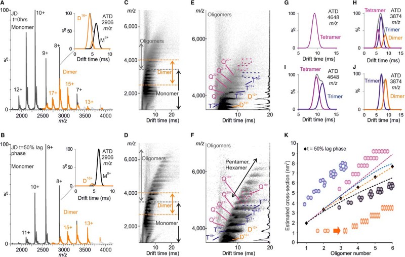
Expansion of polyglutamine stretches leads to the formation of polyglutamine-containing neuronal aggregates and neuronal death in nine diseases for which there currently are no treatments or cures. This is largely due to a lack in understanding of the mechanisms by which expanded polyglutamine regions contribute to aggregation and disease. To complicate matters further, several of the polyglutamine-disease related proteins, including ataxin-3, have a multistage aggregation mechanism in which flanking domain self-assembly precedes polyglutamine aggregation yet is influenced by polyglutamine expansion. How polyglutamine expansion influences flanking domain aggregation is poorly understood. Here, we use a combination of mass spectrometry and biophysical approaches to investigate this issue for ataxin-3. We show that the conformational dynamics of the flanking Josephin domain in ataxin-3 with an expanded polyglutamine tract are altered in comparison to those exhibited by its nonexpanded counterpart, specifically within the aggregation-prone region of the Josephin domain (amino acid residues 73-96). Expansion thus exposes this region more frequently in ataxin-3 containing an expanded polyglutamine tract, providing a molecular explanation of why aggregation is accelerated upon polyglutamine expansion. Here, harnessing the power of ion mobility spectrometry-mass spectrometry, oligomeric species formed during aggregation are characterized and a model for oligomer growth proposed. The results suggest that a conformational change occurs at the dimer level that initiates self-assembly. New insights into ataxin-3 fibril architecture are also described, revealing the region of the Josephin domain involved in protofibril formation and demonstrating that polyglutamine aggregation proceeds as a distinct second step after protofibril formation without requiring structural rearrangement of the protofibril core. Overall, the results enable the effect of polyglutamine expansion on every stage of ataxin-3 self-assembly, from monomer through to fibril, to be described and a rationale for expedited aggregation upon polyglutamine expansion to be provided.
聚谷氨酰胺链的扩展会导致在九种目前尚无治疗方法或治愈手段的疾病中形成含聚谷氨酰胺的神经元聚集体并导致神经元死亡。这主要是由于对扩展的聚谷氨酰胺区域促成聚集体形成和疾病的机制缺乏了解。更复杂的是,几种与聚谷氨酰胺疾病相关的蛋白质,包括ataxin-3,具有多阶段聚集机制,其中侧翼结构域的自组装先于聚谷氨酰胺聚集,但又受聚谷氨酰胺扩展的影响。聚谷氨酰胺扩展如何影响侧翼结构域聚集尚不清楚。在此,我们结合质谱和生物物理方法来研究ataxin-3的这个问题。我们表明,与无扩展对应物相比,具有扩展聚谷氨酰胺序列的ataxin-3中侧翼约瑟芬结构域的构象动力学发生了改变,特别是在约瑟芬结构域的易聚集区域(氨基酸残基73 - 96)内。因此,扩展使该区域在含有扩展聚谷氨酰胺序列的ataxin-3中更频繁地暴露,这为聚谷氨酰胺扩展时聚集加速提供了分子解释。在此,利用离子淌度质谱的强大功能,对聚集过程中形成的寡聚体种类进行了表征,并提出了寡聚体生长模型。结果表明,在二聚体水平发生构象变化从而启动自组装。还描述了对ataxin-3原纤维结构的新见解,揭示了约瑟芬结构域中参与原纤维形成的区域,并证明聚谷氨酰胺聚集在原纤维形成后作为一个独特的第二步进行,而不需要原纤维核心的结构重排。总体而言,这些结果能够描述聚谷氨酰胺扩展对ataxin-3从单体到原纤维的自组装各个阶段的影响,并为聚谷氨酰胺扩展时加速聚集提供一个基本原理。