Ding Y, Lawrence N, Olié E, Cyprien F, le Bars E, Bonafé A, Phillips M L, Courtet P, Jollant F
McGill Group for Suicide Studies, Douglas Mental Health University Institute, McGill University, Montreal, QC, Canada.
Mood Disorders Centre, School of Psychology, College of Life and Environmental Sciences, University of Exeter, Exeter, UK.
Transl Psychiatry. 2015 Feb 24;5(2):e516. doi: 10.1038/tp.2015.1.
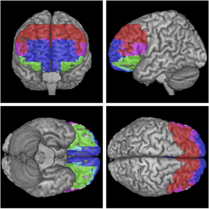
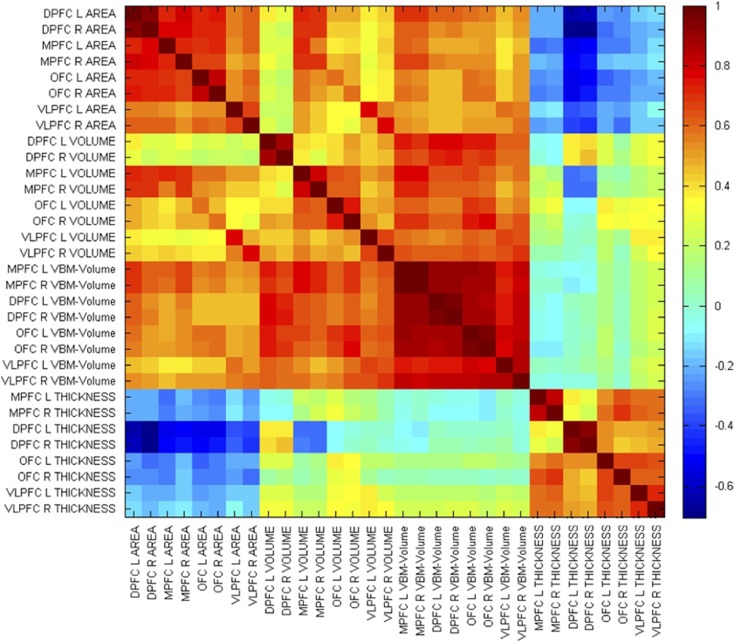
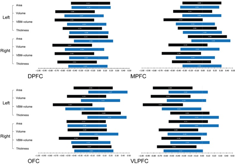
The vulnerability to suicidal behavior has been modeled in deficits in both valuation and cognitive control processes, mediated by ventral and dorsal prefrontal cortices. To uncover potential markers of suicidality based on this model, we measured several brain morphometric parameters using 1.5T magnetic resonance imaging in a large sample and in a specifically designed study. We then tested their classificatory properties. Three groups were compared: euthymic suicide attempters with a past history of mood disorders and suicidal behavior (N=67); patient controls with a past history of mood disorders but not suicidal behavior (N=82); healthy controls without any history of mental disorder (N=82). A hypothesis-driven region-of-interest approach was applied targeting the orbitofrontal cortex (OFC), ventrolateral (VLPFC), dorsal (DPFC) and medial (including anterior cingulate cortex; MPFC) prefrontal cortices. Both voxel-based (SPM8) and surface-based morphometry (Freesurfer) analyses were used to comprehensively evaluate cortical gray matter measure, volume, surface area and thickness. Reduced left VLPFC volume in attempters vs both patient groups was found (P=0.001, surviving multiple comparison correction, Cohen's d=0.65 95% (0.33-0.99) between attempters and healthy controls). In addition, reduced measures in OFC and DPFC, but not MPFC, were found with moderate effect sizes in suicide attempters vs healthy controls (Cohen's d between 0.34 and 0.52). Several of these measures were correlated with suicidal variables. When added to mood disorder history, left VLPFC volume increased within-sample specificity in identifying attempters in a significant but limited way. Our study, therefore, confirms structural prefrontal alterations in individuals with histories of suicide attempts. A future clinical application of these markers will, however, necessitate further research.
自杀行为易感性已被建模为价值评估和认知控制过程的缺陷,由腹侧和背侧前额叶皮层介导。为了基于该模型揭示自杀倾向的潜在标志物,我们在一个大样本中并通过一项专门设计的研究,使用1.5T磁共振成像测量了几个脑形态计量学参数。然后我们测试了它们的分类属性。比较了三组:有情绪障碍和自杀行为病史的心境正常的自杀未遂者(N = 67);有情绪障碍病史但无自杀行为的患者对照(N = 82);无任何精神障碍病史的健康对照(N = 82)。采用假设驱动的感兴趣区域方法,针对眶额皮质(OFC)、腹外侧(VLPFC)、背侧(DPFC)和内侧(包括前扣带回皮质;MPFC)前额叶皮层。基于体素的(SPM8)和基于表面的形态计量学(Freesurfer)分析均被用于全面评估皮质灰质测量值、体积、表面积和厚度。发现与两个患者组相比,未遂者的左侧VLPFC体积减小(P = 0.001,在多重比较校正后存活,未遂者与健康对照之间的Cohen's d = 0.65,95%(0.33 - 0.99))。此外,在自杀未遂者与健康对照中,发现OFC和DPFC的测量值减小,但MPFC未减小,效应大小适中(Cohen's d在0.34至0.52之间)。这些测量值中的几个与自杀变量相关。当加入情绪障碍病史时,左侧VLPFC体积在识别未遂者方面以显著但有限的方式增加了样本内特异性。因此,我们的研究证实了有自杀未遂史个体的前额叶结构改变。然而,这些标志物未来的临床应用需要进一步研究。