• 文献检索
  • 文档翻译
  • 深度研究
  • 学术资讯
  • Suppr Zotero 插件Zotero 插件
  • 邀请有礼
  • 套餐&价格
  • 历史记录
应用&插件
Suppr Zotero 插件Zotero 插件浏览器插件Mac 客户端Windows 客户端微信小程序
定价
高级版会员购买积分包购买API积分包
服务
文献检索文档翻译深度研究API 文档MCP 服务
关于我们
关于 Suppr公司介绍联系我们用户协议隐私条款
关注我们

Suppr 超能文献

核心技术专利:CN118964589B侵权必究
粤ICP备2023148730 号-1Suppr @ 2026

文献检索

告别复杂PubMed语法,用中文像聊天一样搜索,搜遍4000万医学文献。AI智能推荐,让科研检索更轻松。

立即免费搜索

文件翻译

保留排版,准确专业,支持PDF/Word/PPT等文件格式,支持 12+语言互译。

免费翻译文档

深度研究

AI帮你快速写综述,25分钟生成高质量综述,智能提取关键信息,辅助科研写作。

立即免费体验

土耳其人群下颌前磨牙根管系统的锥形束计算机断层扫描研究:确定根管口形状的理论模型

A cone-beam computed tomographic study of root canal systems in mandibular premolars in a Turkish population: Theoretical model for determining orifice shape.

作者信息

Arslan Hakan, Capar Ismail Davut, Ertas Elif Tarim, Ertas Huseyin, Akcay Merve

机构信息

Department of Endodontics, Faculty of Dentistry, Ataturk University, Erzurum, Turkiye.

Department of Endodontics, Faculty of Dentistry, Izmir Katip Celebi University, Izmir, Turkiye.

出版信息

Eur J Dent. 2015 Jan-Mar;9(1):11-19. doi: 10.4103/1305-7456.149632.

DOI:10.4103/1305-7456.149632
PMID:25713478
原文链接:https://pmc.ncbi.nlm.nih.gov/articles/PMC4319286/
Abstract

OBJECTIVE

The purposes of this retrospective study were to represent a newly designed theoretical model for determining orifice shape and morphologic properties of mandibular premolars and to correlate these findings with each other.

MATERIALS AND METHODS

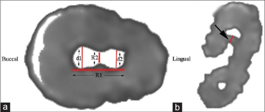
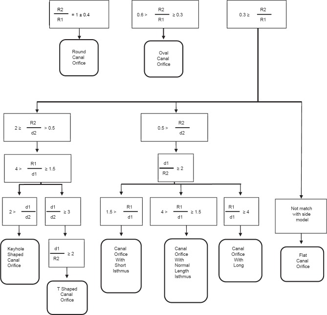
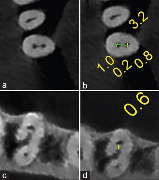
A total of 287 mandibular premolar images obtained from 88 patients by cone-beam computed tomography were included in this study. The measurements were performed below the cementoenamel junction, and different orifice configurations were defined in accordance with various ratios. The age and gender of the patient, the tooth type and position, the number of roots, orifice configuration, root canal configuration, presence of C-shaped canal, and the presence of radicular groove were recorded. It was also recorded whether the root canal becomes round or not and if any, length of the root canal from the orifice to the section in which it becomes round. Furthermore, the theoretical model for determining orifice shape was defined after measurements. The orifice shape was determined as round, oval, flat, keyhole-shaped, and T-shaped, and orifices with short, normal length, and long isthmus. Statistical analyses were performed using Chi-square and Spearman's rank correlation tests (P = 0.05).

RESULTS

Orifice configurations were, usually, flat (37%), or keyhole-shaped (23%). The prevalence of T-shaped was found to be 3.8%. The prevalence of C-shaped canals was found to be 2.1%. The percentage of root canals that became round in the middle or apical thirds was 95.1%. Radicular grooves were detected in 37 (24%) of first premolars and six (4.5%) of second premolars. Statistical analysis revealed that the mean length of distance until the canal reached a round shape varied according to age group (r = -0.270; P < 0.001). There was a statistically significant difference between radicular groove and tooth type (P < 0.001).

CONCLUSIONS

The mean length of distance until the canal reached a round shape correlated with the patient's age. The new theoretical model could be beneficial to determine orifice configurations.

摘要

目的

本回顾性研究的目的是提出一种新设计的理论模型,用于确定下颌前磨牙的根管口形状和形态学特征,并将这些发现相互关联。

材料与方法

本研究纳入了通过锥形束计算机断层扫描从88例患者获得的287张下颌前磨牙图像。测量在牙骨质牙釉质界下方进行,并根据不同比例定义不同的根管口形态。记录患者的年龄和性别、牙齿类型和位置、牙根数量、根管口形态、根管形态、C形根管的存在情况以及根面沟的存在情况。还记录根管是否变圆,以及如果变圆,从根管口到其变圆处的根管长度。此外,在测量后定义了确定根管口形状的理论模型。根管口形状确定为圆形、椭圆形、扁平形、钥匙孔形和T形,以及峡部短、正常长度和长的根管口。使用卡方检验和Spearman等级相关性检验进行统计分析(P = 0.05)。

结果

根管口形态通常为扁平形(37%)或钥匙孔形(23%)。发现T形的患病率为3.8%。发现C形根管的患病率为2.1%。在根管中三分之一或根尖三分之一处变圆的根管百分比为95.1%。在37颗(24%)第一前磨牙和6颗(4.5%)第二前磨牙中检测到根面沟。统计分析显示,直到根管变圆的平均距离长度根据年龄组而变化(r = -0.270;P < 0.001)。根面沟与牙齿类型之间存在统计学显著差异(P < 0.001)。

结论

直到根管变圆的平均距离长度与患者年龄相关。新的理论模型可能有助于确定根管口形态。

https://cdn.ncbi.nlm.nih.gov/pmc/blobs/d890/4319286/fd2bf0ce145b/EJD-9-11-g003.jpg
https://cdn.ncbi.nlm.nih.gov/pmc/blobs/d890/4319286/9ba3732b4ebe/EJD-9-11-g001.jpg
https://cdn.ncbi.nlm.nih.gov/pmc/blobs/d890/4319286/3383a94b23bd/EJD-9-11-g002.jpg
https://cdn.ncbi.nlm.nih.gov/pmc/blobs/d890/4319286/fd2bf0ce145b/EJD-9-11-g003.jpg
https://cdn.ncbi.nlm.nih.gov/pmc/blobs/d890/4319286/9ba3732b4ebe/EJD-9-11-g001.jpg
https://cdn.ncbi.nlm.nih.gov/pmc/blobs/d890/4319286/3383a94b23bd/EJD-9-11-g002.jpg
https://cdn.ncbi.nlm.nih.gov/pmc/blobs/d890/4319286/fd2bf0ce145b/EJD-9-11-g003.jpg

相似文献

1
A cone-beam computed tomographic study of root canal systems in mandibular premolars in a Turkish population: Theoretical model for determining orifice shape.土耳其人群下颌前磨牙根管系统的锥形束计算机断层扫描研究:确定根管口形状的理论模型
Eur J Dent. 2015 Jan-Mar;9(1):11-19. doi: 10.4103/1305-7456.149632.
2
Cone-beam Computed Tomographic Analysis of C-shaped Canals and Radicular Grooves in Mandibular Premolars: Prevalence and Related Factors.下颌前磨牙C形根管和根管沟的锥束计算机断层扫描分析:患病率及相关因素
J Contemp Dent Pract. 2019 Nov 1;20(11):1350-1354.
3
Cone-beam computed tomography evaluation of C-shaped root and canal morphology of mandibular premolars.下颌前磨牙 C 形根管和根管形态的锥形束 CT 评估。
BMC Oral Health. 2021 May 4;21(1):236. doi: 10.1186/s12903-021-01596-y.
4
Investigation using cone beam computed tomography analysis, of radicular grooves and canal configurations of mandibular premolars in a Turkish subpopulation.利用锥形束计算机断层扫描分析,研究土耳其人群下颌前磨牙的根管形态和根管侧支。
Arch Oral Biol. 2019 Nov;107:104517. doi: 10.1016/j.archoralbio.2019.104517. Epub 2019 Aug 10.
5
A cone-beam computed tomography study of C-shaped root canal systems in mandibular second premolars in a Taiwan Chinese subpopulation.一项针对中国台湾人群下颌第二前磨牙 C 型根管系统的锥形束 CT 研究。
J Formos Med Assoc. 2018 Dec;117(12):1086-1092. doi: 10.1016/j.jfma.2017.12.001. Epub 2018 Jan 12.
6
Use of cone-beam computed tomography to evaluate C-shaped root canal systems in mandibular second molars in a Turkish subpopulation: a retrospective study.使用锥形束计算机断层扫描评估土耳其亚人群下颌第二磨牙的C形根管系统:一项回顾性研究。
Int Endod J. 2013 Nov;46(11):1032-8. doi: 10.1111/iej.12094. Epub 2013 Mar 22.
7
A cone-beam computed tomography study of root canal morphology of maxillary and mandibular premolars in a Turkish population.土耳其人群上颌和下颌前磨牙根管形态的锥形束计算机断层扫描研究
Acta Odontol Scand. 2014 Nov;72(8):701-6. doi: 10.3109/00016357.2014.898091. Epub 2014 May 15.
8
Cone beam computed tomography evaluation of c-shaped canal morphology in mandibular premolar teeth.锥形束计算机断层扫描评估下颌前磨牙C形根管形态
Eur Oral Res. 2024 May 5;58(2):70-75. doi: 10.26650/eor.20241175997.
9
Prevalence and morphology of multiple roots, root canals and C-shaped canals in mandibular premolars from cone-beam computed tomography images in a Thai population.泰国人群下颌前磨牙多根、根管及C形根管的患病率和形态:基于锥形束计算机断层扫描图像的研究
J Dent Sci. 2021 Jan;16(1):201-207. doi: 10.1016/j.jds.2020.06.010. Epub 2020 Jun 18.
10
Frequency of non-single canals in mandibular premolars and correlations with other anatomical variants: an in vivo cone beam computed tomography study.下颌前磨牙非单根管的频率及其与其他解剖变异的相关性:一项体内锥形束 CT 研究。
BMC Oral Health. 2019 Dec 4;19(1):272. doi: 10.1186/s12903-019-0972-5.

引用本文的文献

1
Prevalence of Isthmi and Root Canal Configurations in Mandibular Permanent Teeth Using Cone-Beam Computed Tomography.下颌恒磨牙应用锥形束 CT 研究其根管和峡部的解剖形态。
Biomed Res Int. 2024 Jul 31;2024:9969860. doi: 10.1155/2024/9969860. eCollection 2024.
2
Systematic review and meta-analysis of root morphology and canal configuration of permanent premolars using cone-beam computed tomography.使用锥形束计算机断层扫描对恒前磨牙的牙根形态和根管形态进行系统评价和荟萃分析。
BMC Oral Health. 2024 Jun 4;24(1):656. doi: 10.1186/s12903-024-04419-y.
3
Roots and Root Canals Characterization of Permanent Mandibular Premolars Analyzed Using the Cone Beam and Micro Computed Tomography-A Systematic Review and Metanalysis.

本文引用的文献

1
A cone-beam computed tomography study of root canal morphology of maxillary and mandibular premolars in a Turkish population.土耳其人群上颌和下颌前磨牙根管形态的锥形束计算机断层扫描研究
Acta Odontol Scand. 2014 Nov;72(8):701-6. doi: 10.3109/00016357.2014.898091. Epub 2014 May 15.
2
Micro-computed tomography study of oval-shaped canals prepared with the self-adjusting file, Reciproc, WaveOne, and ProTaper universal systems.使用自调式锉、Reciproc、WaveOne和ProTaper通用系统制备的椭圆形根管的微计算机断层扫描研究。
J Endod. 2013 Aug;39(8):1060-6. doi: 10.1016/j.joen.2013.04.009. Epub 2013 May 22.
3
A cone-beam computed tomography study of the root canal morphology of mandibular first premolars and the location of root canal orifices and apical foramina in a Chinese subpopulation.
使用锥形束和微型计算机断层扫描分析的下颌恒前磨牙牙根及根管特征:一项系统评价与Meta分析
J Clin Med. 2023 Mar 11;12(6):2183. doi: 10.3390/jcm12062183.
4
Endodontic Treatment of C-shaped Mandibular Premolars: A Case Report and Review of Literature.下颌C形前磨牙的根管治疗:病例报告及文献综述
Iran Endod J. 2021 Fall;16(4):244-253. doi: 10.22037/iej.v16i4.34187.
5
Canal configuration and root morphology of mandibular premolars using cone-beam computed tomography in a Korean population.利用锥形束 CT 研究韩国人群下颌前磨牙根管形态和根尖孔位置。
Clin Oral Investig. 2022 Mar;26(3):3325-3332. doi: 10.1007/s00784-021-04313-9. Epub 2021 Nov 30.
6
Root canal morphology of the mandibular second premolar: a systematic review and meta-analysis.下颌第二前磨牙根管形态:系统评价和荟萃分析。
BMC Oral Health. 2021 Jun 16;21(1):309. doi: 10.1186/s12903-021-01668-z.
7
Root and Canal Morphology of Mandibular Premolar Teeth in a Kuwaiti Subpopulation: A CBCT Clinical Study.科威特人群下颌前磨牙的根管和牙根形态:CBCT 临床研究。
Eur Endod J. 2020 Dec;5(3):248-256. doi: 10.14744/eej.2020.40085.
8
The maxillary molar endodontic access opening: A microscope-based approach.上颌磨牙牙髓治疗的开髓:基于显微镜的方法。
Eur J Dent. 2016 Jul-Sep;10(3):439-446. doi: 10.4103/1305-7456.184153.
一项关于中国人群下颌第一前磨牙根管形态和根管口及根尖孔位置的锥形束 CT 研究。
J Endod. 2013 Apr;39(4):435-8. doi: 10.1016/j.joen.2012.11.003. Epub 2013 Jan 16.
4
Morphology of maxillary first and second molars analyzed by cone-beam computed tomography in a korean population: variations in the number of roots and canals and the incidence of fusion.锥形束计算机断层扫描分析韩国人群上颌第一、二磨牙的形态:牙根和根管数量的变异及融合的发生率。
J Endod. 2012 Aug;38(8):1063-8. doi: 10.1016/j.joen.2012.04.025. Epub 2012 Jun 20.
5
Maxillary first Molar with three canal orifices in MesioBuccal root.上颌第一磨牙近中颊根有三个根管口。
J Conserv Dent. 2011 Oct;14(4):436-7. doi: 10.4103/0972-0707.87222.
6
Use of cone-beam computed tomography to evaluate root and canal morphology of mandibular molars in Chinese individuals.应用锥形束 CT 评估中国人下颌磨牙的根管及牙根形态。
Int Endod J. 2011 Nov;44(11):990-9. doi: 10.1111/j.1365-2591.2011.01904.x. Epub 2011 Jun 10.
7
C-shaped root canal system in mandibular second molars in a Chinese population evaluated by cone-beam computed tomography.锥形束计算机断层扫描评估的中国人下颌第二磨牙的 C 型根管系统。
Int Endod J. 2011 Sep;44(9):857-62. doi: 10.1111/j.1365-2591.2011.01896.x. Epub 2011 May 21.
8
Use of CBCT to identify the morphology of maxillary permanent molar teeth in a Chinese subpopulation.应用 CBCT 分析中国人群上颌恒磨牙的解剖形态。
Int Endod J. 2011 Feb;44(2):162-9. doi: 10.1111/j.1365-2591.2010.01826.x. Epub 2010 Nov 22.
9
Palatogingival groove: endodontic-periodontal management--case report.腭龈沟:牙髓-牙周联合治疗--病例报告。
J Endod. 2010 Oct;36(10):1717-20. doi: 10.1016/j.joen.2010.06.025. Epub 2010 Aug 17.
10
Cone-beam computed tomography study of root and canal morphology of maxillary first and second molars in an Indian population.上颌第一、二磨牙根管及形态的锥形束 CT 研究。
J Endod. 2010 Oct;36(10):1622-7. doi: 10.1016/j.joen.2010.07.006. Epub 2010 Aug 30.