Kawashiri Masa-Aki, Nakanishi Chiaki, Tsubokawa Toshinari, Shimojima Masaya, Yoshida Shohei, Yoshimuta Tsuyoshi, Konno Tetsuo, Yamagishi Masakazu, Hayashi Kenshi
Division of Cardiovascular Medicine, Kanazawa University Graduate School of Medicine, Kanazawa, Japan.
J Cardiovasc Pharmacol. 2015 Jun;65(6):601-6. doi: 10.1097/FJC.0000000000000231.
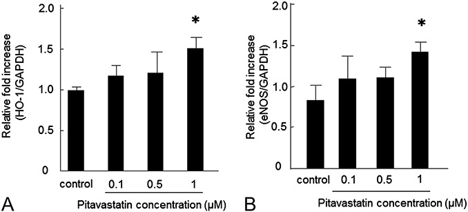
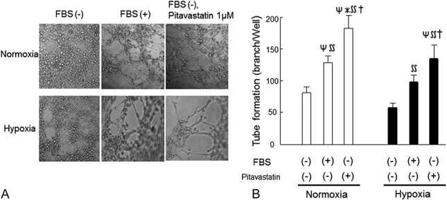
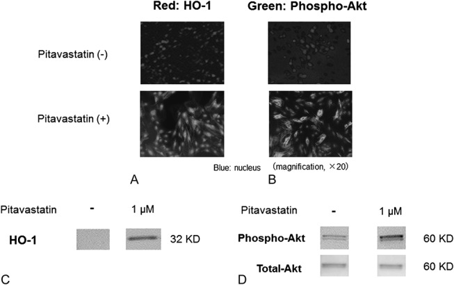
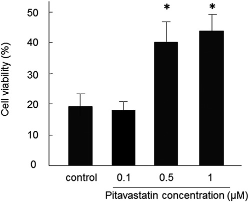
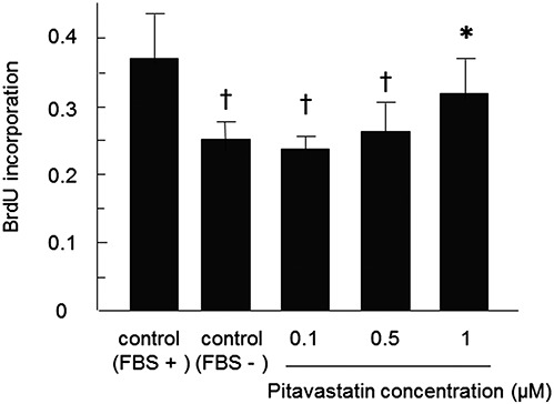
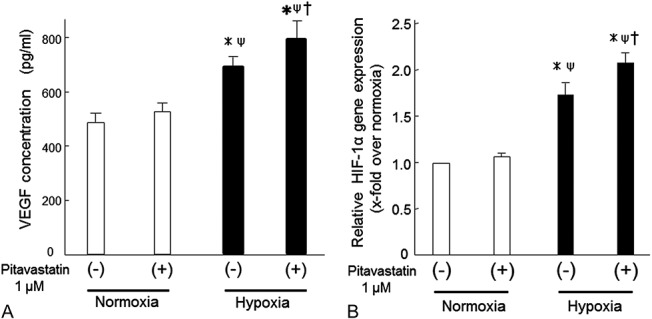
Although mesenchymal stem cells (MSCs) have a therapeutic potential for the repair of tissue injuries, their poor viability in damaged tissue limits their effectiveness. Statins can induce an increased production of heme oxygenase-1 (HO-1), which may prevent this detrimental effect in MSCs. We investigated the protective effect of statin-induced overexpression of HO-1 by examining changes in gene expression and function in MSCs after pitavastatin treatment. The relative expression of the HO-1 and endothelial nitric oxide synthase genes in MSCs was significantly increased after treatment with pitavastatin (MSCs). Immunocytological analysis showed that MSCs also stained with phospho-Akt. After exposure to oxidative stress, MSCs showed increased resistance to induced cell death compared with control MSCs. Under serum starvation conditions, MSCs treated with 1 μM pitavastatin showed enhanced cell proliferation and a marked increase in vascular endothelial growth factor production compared with control MSCs. Interestingly, MSCs showed enhanced tube formation under both normoxia and hypoxia. These results demonstrate that pitavastatin can enhance endogenous HO-1 expression in MSCs, which may protect the cells into the environment of oxidative stress with partial activation of endothelial nitric oxide synthase and Akt phosphorylation.
尽管间充质干细胞(MSCs)具有修复组织损伤的治疗潜力,但其在受损组织中的低存活率限制了它们的有效性。他汀类药物可诱导血红素加氧酶-1(HO-1)产量增加,这可能会防止MSCs出现这种有害影响。我们通过检测匹伐他汀处理后MSCs的基因表达和功能变化,研究了他汀类药物诱导的HO-1过表达的保护作用。匹伐他汀处理后,MSCs中HO-1和内皮型一氧化氮合酶基因的相对表达显著增加(MSCs)。免疫细胞分析表明,MSCs也用磷酸化Akt染色。暴露于氧化应激后,与对照MSCs相比,MSCs对诱导的细胞死亡表现出更高的抗性。在血清饥饿条件下,与对照MSCs相比,用1μM匹伐他汀处理的MSCs显示出增强的细胞增殖和血管内皮生长因子产量的显著增加。有趣的是,MSCs在常氧和低氧条件下均显示出增强的管形成。这些结果表明,匹伐他汀可增强MSCs中内源性HO-1的表达,这可能通过部分激活内皮型一氧化氮合酶和Akt磷酸化来保护细胞免受氧化应激环境的影响。