Dinh Minh H, Anderson Meegan R, McRaven Michael D, Cianci Gianguido C, McCoombe Scott G, Kelley Z L, Gioia Casey J, Fought Angela J, Rademaker Alfred W, Veazey Ronald S, Hope Thomas J
Division of Infectious Diseases, Department of Medicine, Northwestern University Feinberg School of Medicine, Chicago, Illinois, United States of America.
Department of Cell & Molecular Biology, Northwestern University Feinberg School of Medicine, Chicago, Illinois, United States of America.
PLoS Pathog. 2015 Mar 6;11(3):e1004729. doi: 10.1371/journal.ppat.1004729. eCollection 2015 Mar.
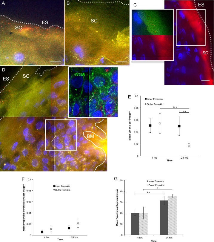
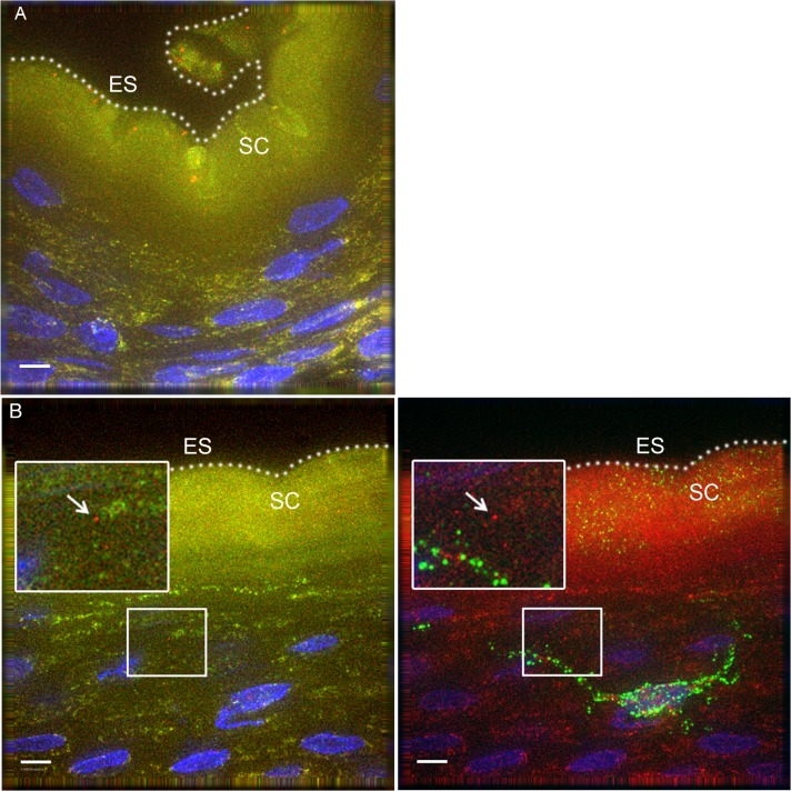
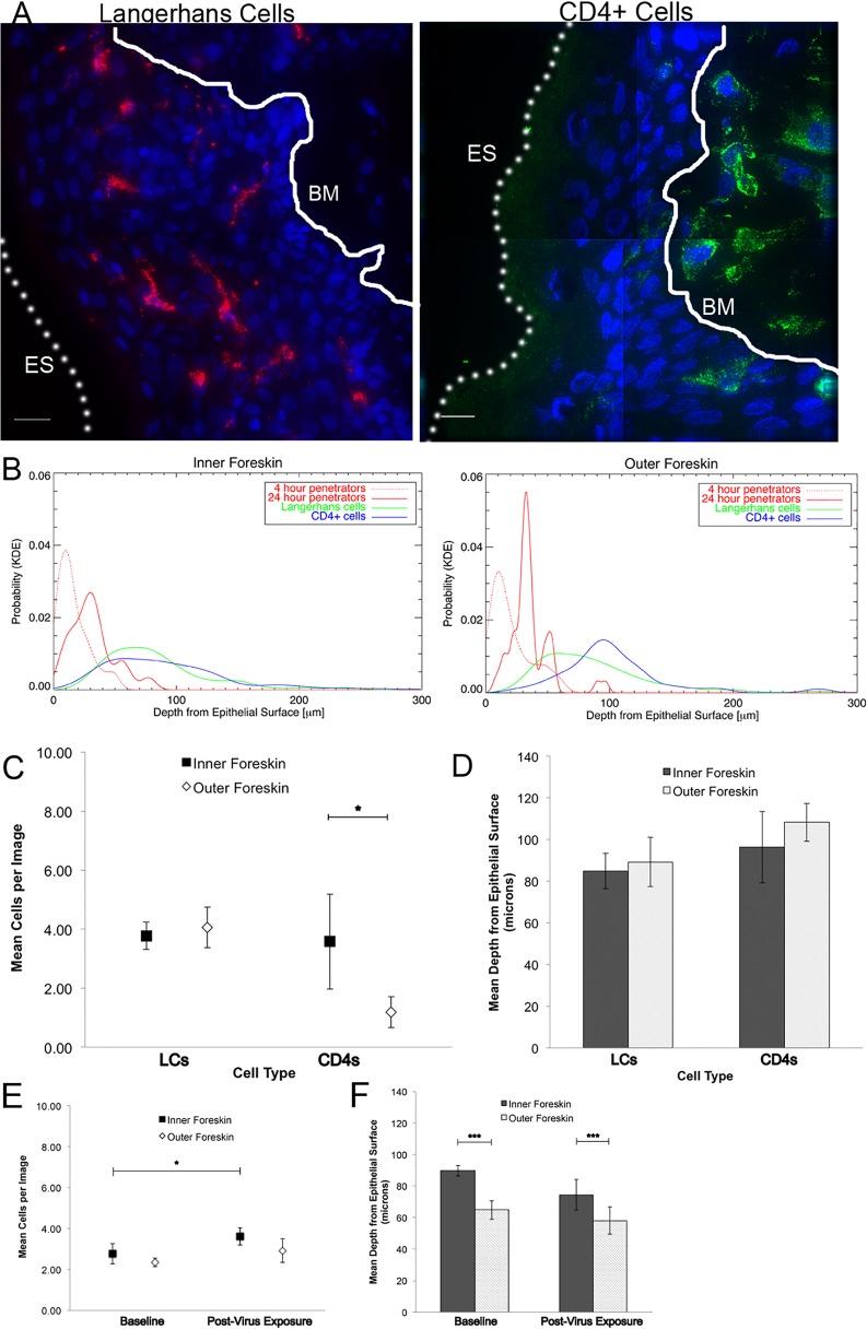
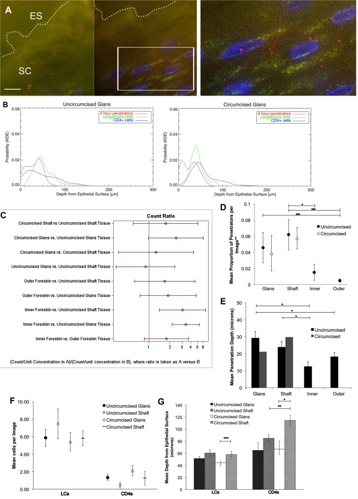
To gain insight into female-to-male HIV sexual transmission and how male circumcision protects against this mode of transmission, we visualized HIV-1 interactions with foreskin and penile tissues in ex vivo tissue culture and in vivo rhesus macaque models utilizing epifluorescent microscopy. 12 foreskin and 14 cadaveric penile specimens were cultured with R5-tropic photoactivatable (PA)-GFP HIV-1 for 4 or 24 hours. Tissue cryosections were immunofluorescently imaged for epithelial and immune cell markers. Images were analyzed for total virions, proportion of penetrators, depth of virion penetration, as well as immune cell counts and depths in the tissue. We visualized individual PA virions breaching penile epithelial surfaces in the explant and macaque model. Using kernel density estimated probabilities of localizing a virion or immune cell at certain tissue depths revealed that interactions between virions and cells were more likely to occur in the inner foreskin or glans penis (from local or cadaveric donors, respectively). Using statistical models to account for repeated measures and zero-inflated datasets, we found no difference in total virions visualized at 4 hours between inner and outer foreskins from local donors. At 24 hours, there were more virions in inner as compared to outer foreskin (0.0495 +/- 0.0154 and 0.0171 +/- 0.0038 virions/image, p = 0.001). In the cadaveric specimens, we observed more virions in inner foreskin (0.0507 +/- 0.0079 virions/image) than glans tissue (0.0167 +/- 0.0033 virions/image, p<0.001), but a greater proportion was seen penetrating uncircumcised glans tissue (0.0458 +/- 0.0188 vs. 0.0151 +/- 0.0100 virions/image, p = 0.099) and to significantly greater mean depths (29.162 +/- 3.908 vs. 12.466 +/- 2.985 μm). Our in vivo macaque model confirmed that virions can breach penile squamous epithelia in a living model. In summary, these results suggest that the inner foreskin and glans epithelia may be important sites for HIV transmission in uncircumcised men.
为深入了解女性到男性的HIV性传播以及男性包皮环切术如何预防这种传播方式,我们利用落射荧光显微镜,在离体组织培养和体内恒河猴模型中观察了HIV-1与包皮和阴茎组织的相互作用。将12个包皮标本和14个尸体阴茎标本与R5嗜性光激活(PA)-GFP HIV-1共培养4或24小时。对组织冰冻切片进行上皮细胞和免疫细胞标志物的免疫荧光成像。分析图像中的病毒体总数、穿透者比例、病毒体穿透深度,以及组织中的免疫细胞计数和深度。我们在离体标本和猕猴模型中观察到单个PA病毒体突破阴茎上皮表面。使用核密度估计法确定病毒体或免疫细胞在特定组织深度定位的概率,结果显示病毒体与细胞之间的相互作用更可能发生在包皮内层或阴茎头(分别来自本地或尸体供体)。使用统计模型来处理重复测量和零膨胀数据集,我们发现本地供体的包皮内层和外层在4小时时观察到的病毒体总数没有差异。在24小时时,包皮内层的病毒体比外层更多(0.0495±0.0154和0.0171±0.0038个病毒体/图像,p = 0.001)。在尸体标本中,我们观察到包皮内层的病毒体(0.0507±0.0079个病毒体/图像)比阴茎头组织(0.0167±0.0033个病毒体/图像,p<0.001)更多,但未环切阴茎头组织中观察到的穿透病毒体比例更高(0.0458±0.0188与0.0151±0.0100个病毒体/图像,p = (此处原文可能有误,推测应为p = 0.099)),且平均穿透深度显著更深(29.162±3.908与12.466±2.985μm)。我们的体内猕猴模型证实病毒体可以在活体模型中突破阴茎鳞状上皮。总之,这些结果表明包皮内层和阴茎头上皮可能是未环切男性中HIV传播的重要部位。