Celikten Berkan, Uzuntas Ceren Feriha, Gulsahi Kamran
Department of Endodontics, Faculty of Dentistry, Ankara University, Ankara, Turkey.
Department of Endodontics, Faculty of Dentistry, Başkent University, Ankara, Turkey.
Biomed Res Int. 2015;2015:591031. doi: 10.1155/2015/591031. Epub 2015 Feb 8.
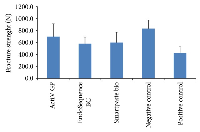
The aim of this study was to compare the vertical fracture resistance of roots obturated with different root canal filling materials and sealers. Crowns of 55 extracted mandibular premolar teeth were removed to provide root lengths of 13 mm. Five roots were saved as negative control group (canals unprepared and unfilled). Fifty root canals were instrumented and then five roots were saved as positive control group (canals prepared but unfilled). The remaining 45 roots were randomly divided into three experimental groups (n = 15 root/group) and obturated with the following procedures: in group 1, glass ionomer-based sealer and cone (ActiV GP obturation system); in group 2, bioceramic sealer and cone (EndoSequence BC obturation system); and in group 3, roots were filled with bioceramic sealer and cone (Smartpaste bio obturation system). All specimens were tested in a universal testing machine for measuring fracture resistance. For each root, the force at the time of fracture was recorded in Newtons. The statistical analysis was performed by using Kruskal-Wallis and post hoc test. There were no significant differences between the three experimental groups. The fracture values of three experimental and negative control groups were significantly higher than the positive control group. Within the limitations of this study, all materials increased the fracture resistance of instrumented roots.
本研究的目的是比较用不同根管充填材料和封闭剂充填的牙根的垂直抗折性。去除55颗拔除的下颌前磨牙的牙冠,使牙根长度为13毫米。保留5个牙根作为阴性对照组(根管未预备和未充填)。对50个根管进行预备,然后保留5个牙根作为阳性对照组(根管已预备但未充填)。将其余45个牙根随机分为三个实验组(每组15个牙根),并按以下步骤进行充填:第1组,使用玻璃离子基封闭剂和牙胶尖(ActiV GP充填系统);第2组,使用生物陶瓷封闭剂和牙胶尖(EndoSequence BC充填系统);第3组,使用生物陶瓷封闭剂和牙胶尖(Smartpaste bio充填系统)。所有标本在万能试验机上进行测试以测量抗折性。对于每个牙根,记录骨折时的力,单位为牛顿。采用Kruskal-Wallis检验和事后检验进行统计分析。三个实验组之间没有显著差异。三个实验组和阴性对照组的骨折值显著高于阳性对照组。在本研究的局限性内,所有材料均提高了预备后牙根的抗折性。