• 文献检索
  • 文档翻译
  • 深度研究
  • 学术资讯
  • Suppr Zotero 插件Zotero 插件
  • 邀请有礼
  • 套餐&价格
  • 历史记录
应用&插件
Suppr Zotero 插件Zotero 插件浏览器插件Mac 客户端Windows 客户端微信小程序
定价
高级版会员购买积分包购买API积分包
服务
文献检索文档翻译深度研究API 文档MCP 服务
关于我们
关于 Suppr公司介绍联系我们用户协议隐私条款
关注我们

Suppr 超能文献

核心技术专利:CN118964589B侵权必究
粤ICP备2023148730 号-1Suppr @ 2026

文献检索

告别复杂PubMed语法,用中文像聊天一样搜索,搜遍4000万医学文献。AI智能推荐,让科研检索更轻松。

立即免费搜索

文件翻译

保留排版,准确专业,支持PDF/Word/PPT等文件格式,支持 12+语言互译。

免费翻译文档

深度研究

AI帮你快速写综述,25分钟生成高质量综述,智能提取关键信息,辅助科研写作。

立即免费体验

人类外周神经对X光成像敏感吗?

Are human peripheral nerves sensitive to X-ray imaging?

作者信息

Scopel Jonas Francisco, de Souza Queiroz Luciano, O'Dowd Francis Pierce, Júnior Marcondes Cavalcante França, Nucci Anamarli, Hönnicke Marcelo Gonçalves

机构信息

Instituto de Ciências da Saúde, Universidade Federal de Goiás, Jataí, Goiás, 75804-020, Brazil.

Departamento de Anatomia Patológica, Universidade Estadual de Campinas, Campinas, São Paulo, 13083-887, Brazil.

出版信息

PLoS One. 2015 Mar 10;10(3):e0116831. doi: 10.1371/journal.pone.0116831. eCollection 2015.

DOI:10.1371/journal.pone.0116831
PMID:25757086
原文链接:https://pmc.ncbi.nlm.nih.gov/articles/PMC4355589/
Abstract

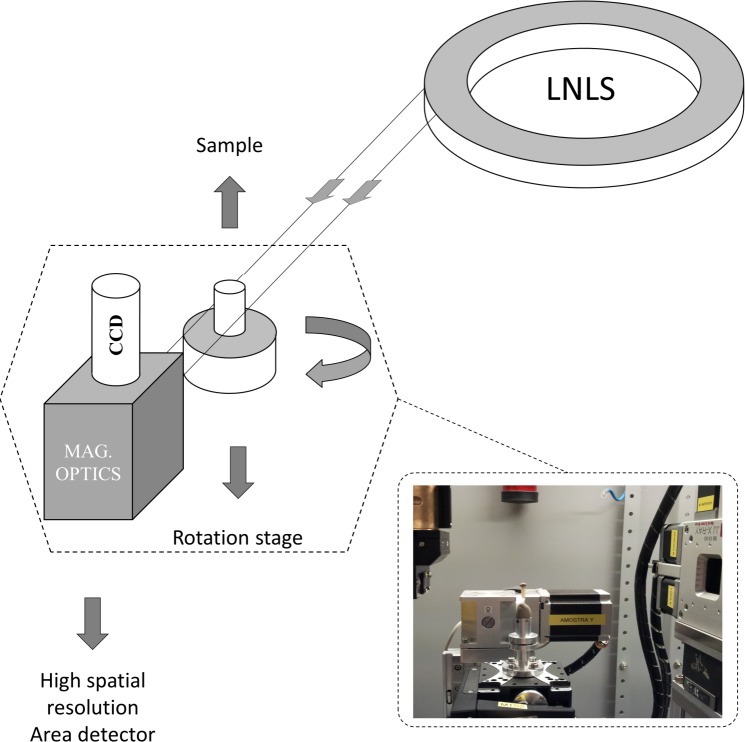
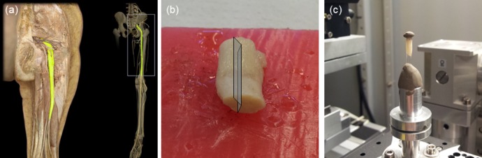
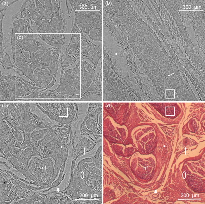
Diagnostic imaging techniques play an important role in assessing the exact location, cause, and extent of a nerve lesion, thus allowing clinicians to diagnose and manage more effectively a variety of pathological conditions, such as entrapment syndromes, traumatic injuries, and space-occupying lesions. Ultrasound and nuclear magnetic resonance imaging are becoming useful methods for this purpose, but they still lack spatial resolution. In this regard, recent phase contrast x-ray imaging experiments of peripheral nerve allowed the visualization of each nerve fiber surrounded by its myelin sheath as clearly as optical microscopy. In the present study, we attempted to produce high-resolution x-ray phase contrast images of a human sciatic nerve by using synchrotron radiation propagation-based imaging. The images showed high contrast and high spatial resolution, allowing clear identification of each fascicle structure and surrounding connective tissue. The outstanding result is the detection of such structures by phase contrast x-ray tomography of a thick human sciatic nerve section. This may further enable the identification of diverse pathological patterns, such as Wallerian degeneration, hypertrophic neuropathy, inflammatory infiltration, leprosy neuropathy and amyloid deposits. To the best of our knowledge, this is the first successful phase contrast x-ray imaging experiment of a human peripheral nerve sample. Our long-term goal is to develop peripheral nerve imaging methods that could supersede biopsy procedures.

摘要

诊断成像技术在评估神经病变的确切位置、病因和范围方面发挥着重要作用,从而使临床医生能够更有效地诊断和处理各种病理状况,如卡压综合征、创伤性损伤和占位性病变。超声和核磁共振成像正成为用于此目的的有用方法,但它们仍缺乏空间分辨率。在这方面,最近对外周神经进行的相衬X射线成像实验能够像光学显微镜一样清晰地显示被髓鞘包裹的每一根神经纤维。在本研究中,我们试图通过基于同步辐射传播的成像来生成人类坐骨神经的高分辨率X射线相衬图像。这些图像显示出高对比度和高空间分辨率,能够清晰地识别每一束 fascicle 结构和周围的结缔组织。突出的成果是通过对厚的人类坐骨神经切片进行相衬X射线断层扫描检测到了此类结构。这可能进一步有助于识别各种病理模式,如沃勒变性、肥厚性神经病、炎性浸润、麻风性神经病和淀粉样沉积。据我们所知,这是首次对人类外周神经样本成功进行的相衬X射线成像实验。我们的长期目标是开发能够取代活检程序的外周神经成像方法。

https://cdn.ncbi.nlm.nih.gov/pmc/blobs/b9d0/4355589/2b690af00308/pone.0116831.g004.jpg
https://cdn.ncbi.nlm.nih.gov/pmc/blobs/b9d0/4355589/ba964ac50d13/pone.0116831.g001.jpg
https://cdn.ncbi.nlm.nih.gov/pmc/blobs/b9d0/4355589/26955c23aafe/pone.0116831.g002.jpg
https://cdn.ncbi.nlm.nih.gov/pmc/blobs/b9d0/4355589/d9edead47df5/pone.0116831.g003.jpg
https://cdn.ncbi.nlm.nih.gov/pmc/blobs/b9d0/4355589/2b690af00308/pone.0116831.g004.jpg

相似文献

1
Are human peripheral nerves sensitive to X-ray imaging?人类外周神经对X光成像敏感吗?
PLoS One. 2015 Mar 10;10(3):e0116831. doi: 10.1371/journal.pone.0116831. eCollection 2015.
2
High resolution X-ray phase contrast synchrotron imaging of normal and ligation damaged rat sciatic nerves.正常及结扎损伤大鼠坐骨神经的高分辨率X射线相衬同步辐射成像
Microsc Res Tech. 2008 Jun;71(6):443-7. doi: 10.1002/jemt.20571.
3
In-line holography and phase-contrast microtomography with high energy x-rays.高能X射线的在线全息术和相衬显微断层成像
Phys Med Biol. 1999 Mar;44(3):741-9. doi: 10.1088/0031-9155/44/3/016.
4
X-ray high-resolution vascular network imaging.X射线高分辨率血管网络成像。
J Microsc. 2004 Aug;215(Pt 2):139-48. doi: 10.1111/j.0022-2720.2004.01362.x.
5
A computed tomography and magnetic resonance imaging study of the variations of the sciatic nerve branches of the pediatric knee: Implications for peripheral nerve blockade.小儿膝关节坐骨神经分支的 CT 和 MRI 研究:对外周神经阻滞的意义。
Clin Anat. 2019 Sep;32(6):836-850. doi: 10.1002/ca.23412. Epub 2019 Jun 19.
6
Optimization of propagation-based x-ray phase-contrast tomography for breast cancer imaging.用于乳腺癌成像的基于传播的X射线相衬断层扫描的优化
Phys Med Biol. 2017 Mar 21;62(6):2315-2332. doi: 10.1088/1361-6560/aa5d3d. Epub 2017 Jan 31.
7
Dose fractionation in synchrotron radiation x-ray phase micro-tomography.同步辐射X射线相微断层扫描中的剂量分割
Phys Med Biol. 2015 Oct 7;60(19):7543-66. doi: 10.1088/0031-9155/60/19/7543. Epub 2015 Sep 15.
8
Development of a Rabbit Model of Radiation-Induced Sciatic Nerve Injury: In Vivo Evaluation Using T2 Relaxation Time Measurements.
J Comput Assist Tomogr. 2015 Jul-Aug;39(4):613-8. doi: 10.1097/RCT.0000000000000241.
9
Synchrotron tomographic images from human lung adenocarcinoma: Three-dimensional reconstruction and histologic correlations.来自人肺腺癌的同步加速器断层图像:三维重建及组织学相关性
Microsc Res Tech. 2017 Oct;80(10):1141-1148. doi: 10.1002/jemt.22910. Epub 2017 Jul 21.
10
X-ray phase-contrast tomography with a compact laser-driven synchrotron source.采用紧凑型激光驱动同步加速器源的X射线相衬断层扫描术。
Proc Natl Acad Sci U S A. 2015 May 5;112(18):5567-72. doi: 10.1073/pnas.1500938112. Epub 2015 Apr 20.

引用本文的文献

1
Analyzer-based X-ray phase contrast imaging using the forward-diffracted o-beam in a few-millimetre-thick Bragg-case asymmetrically cut crystal.在几毫米厚的布拉格情况非对称切割晶体中,利用前向衍射o光进行基于分析仪的X射线相衬成像。
J Appl Crystallogr. 2025 Apr 25;58(Pt 3):789-795. doi: 10.1107/S1600576725002985. eCollection 2025 Jun 1.
2
Imaging fascicular organization of rat sciatic nerves with fast neural electrical impedance tomography.应用快速神经电阻抗断层成像技术对大鼠坐骨神经束结构进行成像。
Nat Commun. 2020 Dec 7;11(1):6241. doi: 10.1038/s41467-020-20127-x.
3
Three-dimensional architecture of human diabetic peripheral nerves revealed by X-ray phase contrast holographic nanotomography.

本文引用的文献

1
Radiation dose and image quality in K-edge subtraction computed tomography of lung in vivo.体内肺 K 边减影 CT 的辐射剂量与图像质量。
J Synchrotron Radiat. 2014 Nov;21(Pt 6):1305-13. doi: 10.1107/S160057751401697X. Epub 2014 Oct 1.
2
High energy x-ray phase contrast CT using glancing-angle grating interferometers.使用掠角光栅干涉仪的高能 X 射线相衬 CT。
Med Phys. 2014 Feb;41(2):021904. doi: 10.1118/1.4860275.
3
X-ray phase imaging: from synchrotron to hospital.X 射线相衬成像:从同步加速器到医院。
X 射线相衬全息纳米断层成像术揭示的人糖尿病外周神经的三维结构。
Sci Rep. 2020 May 5;10(1):7592. doi: 10.1038/s41598-020-64430-5.
4
Phase-contrast x-ray tomography of neuronal tissue at laboratory sources with submicron resolution.实验室光源下具有亚微米分辨率的神经元组织相衬X射线断层扫描术。
J Med Imaging (Bellingham). 2020 Jan;7(1):013502. doi: 10.1117/1.JMI.7.1.013502. Epub 2020 Feb 20.
Philos Trans A Math Phys Eng Sci. 2014 Jan 27;372(2010):20130023. doi: 10.1098/rsta.2013.0023. Print 2014 Mar 6.
4
Source effects in analyzer-based X-ray phase contrast imaging with conventional sources.基于传统光源的分析仪型X射线相衬成像中的光源效应
Rev Sci Instrum. 2012 Nov;83(11):113702. doi: 10.1063/1.4767243.
5
Ultrasound and magnetic resonance imaging of the peripheral nerves: current techniques, promising directions, and open issues.外周神经的超声与磁共振成像:当前技术、前景方向及未决问题
Semin Musculoskelet Radiol. 2010 Nov;14(5):463-72. doi: 10.1055/s-0030-1268067. Epub 2010 Nov 11.
6
Imaging of the peripheral nerves.周围神经成像
Semin Musculoskelet Radiol. 2010 Nov;14(5):461-2. doi: 10.1055/s-0030-1268395. Epub 2010 Nov 11.
7
Development of an x-ray prism for analyzer based imaging systems.用于基于分析仪的成像系统的X射线棱镜的开发。
Rev Sci Instrum. 2010 Aug;81(8):085108. doi: 10.1063/1.3478018.
8
Design and implementation of a compact low-dose diffraction enhanced medical imaging system.紧凑型低剂量衍射增强医学成像系统的设计与实现
Acad Radiol. 2009 Aug;16(8):911-7. doi: 10.1016/j.acra.2009.02.007. Epub 2009 Apr 17.
9
High-resolution X-ray tomography of the human inner ear: synchrotron radiation-based study of nerve fibre bundles, membranes and ganglion cells.人类内耳的高分辨率X射线断层扫描:基于同步辐射的神经纤维束、膜和神经节细胞研究。
J Microsc. 2009 Apr;234(1):95-102. doi: 10.1111/j.1365-2818.2009.03143.x.
10
Hard X-ray phase-contrast imaging with the Compact Light Source based on inverse Compton X-rays.基于逆康普顿X射线的紧凑型光源硬X射线相衬成像
J Synchrotron Radiat. 2009 Jan;16(Pt 1):43-7. doi: 10.1107/S090904950803464X. Epub 2008 Nov 27.