Institute of Multidisciplinary Research for Advanced Materials, Tohoku University, , 2-1-1 Katahira, Aoba-ku, Sendai, Miyagi 980-8577, Japan.
Philos Trans A Math Phys Eng Sci. 2014 Jan 27;372(2010):20130023. doi: 10.1098/rsta.2013.0023. Print 2014 Mar 6.
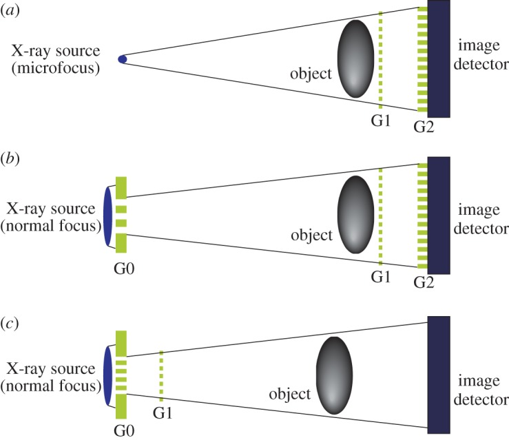
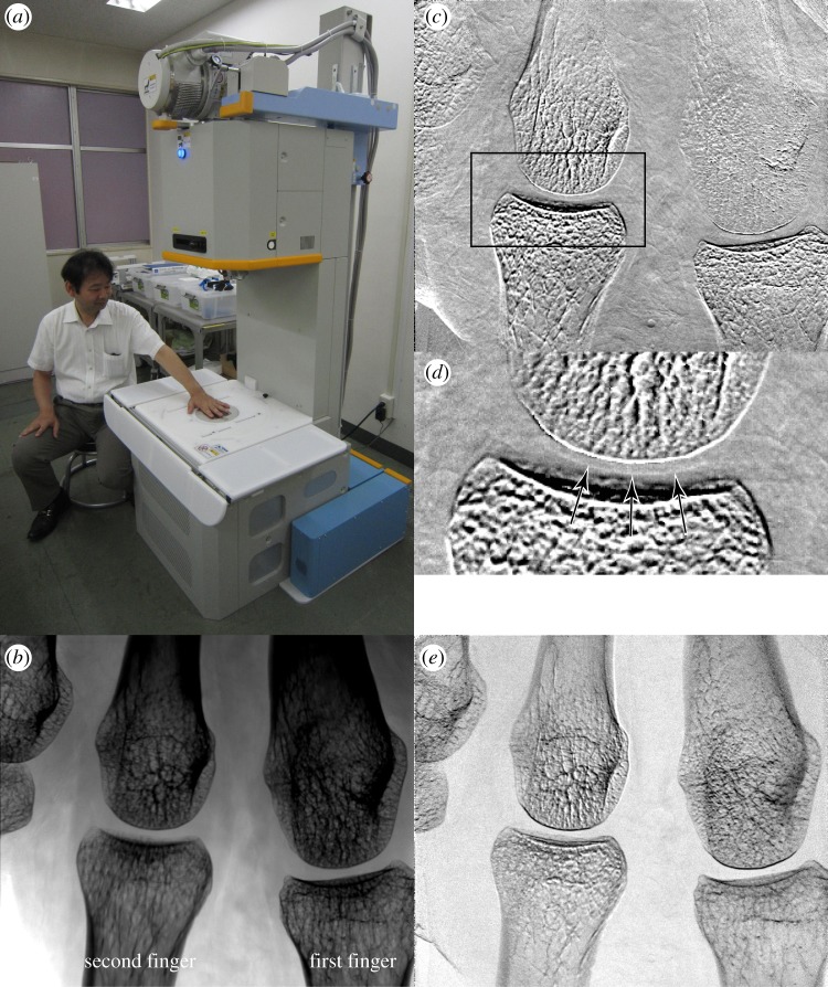
With the aim of clinical applications of X-ray phase imaging based on Talbot-Lau-type grating interferometry to joint diseases and breast cancer, machines employing a conventional X-ray generator have been developed and installed in hospitals. The machine operation especially for diagnosing rheumatoid arthritis is described, which relies on the fact that cartilage in finger joints can be depicted with a dose of several milligray. The palm of a volunteer observed with 19 s exposure (total scan time: 32 s) is reported with a depicted cartilage feature in joints. This machine is now dedicated for clinical research with patients.
为了将基于泰伯-劳型光栅干涉法的 X 射线相位成像技术应用于关节疾病和乳腺癌的临床诊断,已经开发并在医院中安装了使用传统 X 射线发生器的设备。本文介绍了一种专门用于诊断类风湿关节炎的设备操作方法,其原理是可以用几毫戈瑞的剂量来描绘手指关节的软骨。本文报告了对志愿者手掌进行 19 秒曝光(总扫描时间:32 秒)的结果,其中显示了关节处软骨的特征。该设备目前正用于对患者进行临床研究。