• 文献检索
  • 文档翻译
  • 深度研究
  • 学术资讯
  • Suppr Zotero 插件Zotero 插件
  • 邀请有礼
  • 套餐&价格
  • 历史记录
应用&插件
Suppr Zotero 插件Zotero 插件浏览器插件Mac 客户端Windows 客户端微信小程序
定价
高级版会员购买积分包购买API积分包
服务
文献检索文档翻译深度研究API 文档MCP 服务
关于我们
关于 Suppr公司介绍联系我们用户协议隐私条款
关注我们

Suppr 超能文献

核心技术专利:CN118964589B侵权必究
粤ICP备2023148730 号-1Suppr @ 2026

文献检索

告别复杂PubMed语法,用中文像聊天一样搜索,搜遍4000万医学文献。AI智能推荐,让科研检索更轻松。

立即免费搜索

文件翻译

保留排版,准确专业,支持PDF/Word/PPT等文件格式,支持 12+语言互译。

免费翻译文档

深度研究

AI帮你快速写综述,25分钟生成高质量综述,智能提取关键信息,辅助科研写作。

立即免费体验

二氧化硅纳米颗粒通过激活丝裂原活化蛋白激酶/核因子E2相关因子2(MAPK/Nrf2)信号通路和核因子κB信号通路,在体外诱导氧化应激、炎症和内皮功能障碍。

Silica nanoparticles induce oxidative stress, inflammation, and endothelial dysfunction in vitro via activation of the MAPK/Nrf2 pathway and nuclear factor-κB signaling.

作者信息

Guo Caixia, Xia Yinye, Niu Piye, Jiang Lizhen, Duan Junchao, Yu Yang, Zhou Xianqing, Li Yanbo, Sun Zhiwei

机构信息

School of Public Health, Capital Medical University, Beijing, People's Republic of China ; Beijing Key Laboratory of Environmental Toxicology, Capital Medical University, Beijing, People's Republic of China.

出版信息

Int J Nanomedicine. 2015 Feb 20;10:1463-77. doi: 10.2147/IJN.S76114. eCollection 2015.

DOI:10.2147/IJN.S76114
PMID:25759575
原文链接:https://pmc.ncbi.nlm.nih.gov/articles/PMC4345992/
Abstract

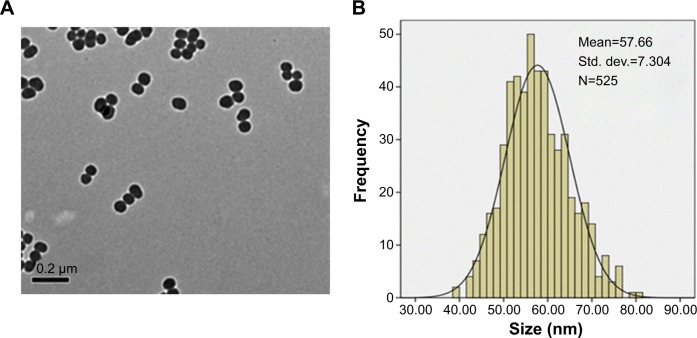
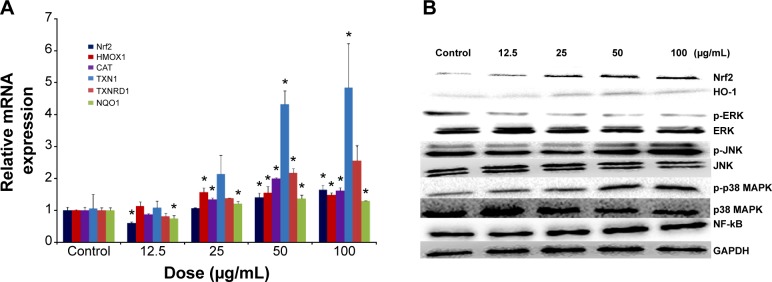
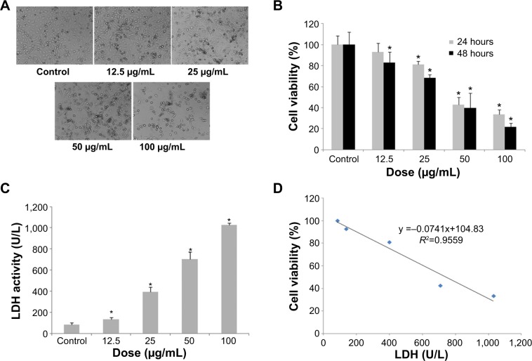
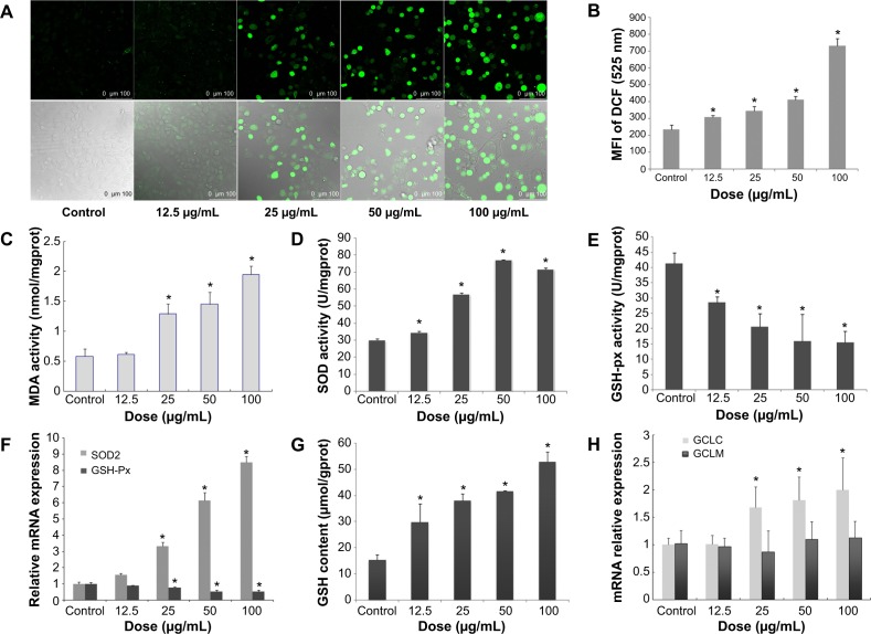
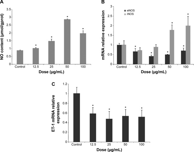
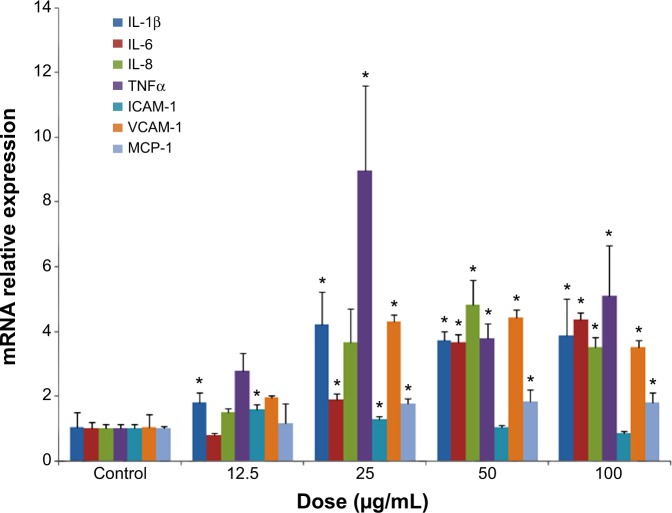
Despite the widespread application of silica nanoparticles (SiNPs) in industrial, commercial, and biomedical fields, their response to human cells has not been fully elucidated. Overall, little is known about the toxicological effects of SiNPs on the cardiovascular system. In this study, SiNPs with a 58 nm diameter were used to study their interaction with human umbilical vein endothelial cells (HUVECs). Dose- and time-dependent decrease in cell viability and damage on cell plasma-membrane integrity showed the cytotoxic potential of the SiNPs. SiNPs were found to induce oxidative stress, as evidenced by the significant elevation of reactive oxygen species generation and malondialdehyde production and downregulated activity in glutathione peroxidase. SiNPs also stimulated release of cytoprotective nitric oxide (NO) and upregulated inducible nitric oxide synthase (NOS) messenger ribonucleic acid, while downregulating endothelial NOS and ET-1 messenger ribonucleic acid, suggesting that SiNPs disturbed the NO/NOS system. SiNP-induced oxidative stress and NO/NOS imbalance resulted in endothelial dysfunction. SiNPs induced inflammation characterized by the upregulation of key inflammatory mediators, including IL-1β, IL-6, IL-8, TNFα, ICAM-1, VCAM-1, and MCP-1. In addition, SiNPs triggered the activation of the Nrf2-mediated antioxidant system, as evidenced by the induction of nuclear factor-κB and MAPK pathway activation. Our findings demonstrated that SiNPs could induce oxidative stress, inflammation, and NO/NOS system imbalance, and eventually lead to endothelial dysfunction via activation of the MAPK/Nrf2 pathway and nuclear factor-κB signaling. This study indicated a potential deleterious effect of SiNPs on the vascular endothelium, which warrants more careful assessment of SiNPs before their application.

摘要

尽管二氧化硅纳米颗粒(SiNPs)在工业、商业和生物医学领域得到了广泛应用,但其对人体细胞的反应尚未完全阐明。总体而言,关于SiNPs对心血管系统的毒理学效应知之甚少。在本研究中,使用直径为58 nm的SiNPs来研究其与人类脐静脉内皮细胞(HUVECs)的相互作用。细胞活力的剂量和时间依赖性降低以及对细胞质膜完整性的损害表明了SiNPs的细胞毒性潜力。发现SiNPs可诱导氧化应激,活性氧生成和丙二醛产生显著升高以及谷胱甘肽过氧化物酶活性下调证明了这一点。SiNPs还刺激了细胞保护性一氧化氮(NO)的释放,并上调了诱导型一氧化氮合酶(NOS)信使核糖核酸,同时下调了内皮型NOS和ET-1信使核糖核酸,这表明SiNPs扰乱了NO/NOS系统。SiNP诱导的氧化应激和NO/NOS失衡导致内皮功能障碍。SiNPs诱导炎症,其特征是关键炎症介质(包括IL-1β、IL-6、IL-8、TNFα、ICAM-1、VCAM-1和MCP-1)上调。此外,SiNPs触发了Nrf2介导的抗氧化系统的激活,核因子-κB的诱导和MAPK途径激活证明了这一点。我们的研究结果表明,SiNPs可诱导氧化应激、炎症和NO/NOS系统失衡,并最终通过激活MAPK/Nrf2途径和核因子-κB信号传导导致内皮功能障碍。本研究表明SiNPs对血管内皮有潜在的有害影响,这在其应用前需要更仔细地评估SiNPs。

https://cdn.ncbi.nlm.nih.gov/pmc/blobs/e343/4345992/945497a35f4d/ijn-10-1463Fig6.jpg
https://cdn.ncbi.nlm.nih.gov/pmc/blobs/e343/4345992/45035dc8c7fd/ijn-10-1463Fig1.jpg
https://cdn.ncbi.nlm.nih.gov/pmc/blobs/e343/4345992/d96f9136d529/ijn-10-1463Fig2.jpg
https://cdn.ncbi.nlm.nih.gov/pmc/blobs/e343/4345992/0feb69ceb559/ijn-10-1463Fig3.jpg
https://cdn.ncbi.nlm.nih.gov/pmc/blobs/e343/4345992/2446d03c88a9/ijn-10-1463Fig4.jpg
https://cdn.ncbi.nlm.nih.gov/pmc/blobs/e343/4345992/5b55988baa8a/ijn-10-1463Fig5.jpg
https://cdn.ncbi.nlm.nih.gov/pmc/blobs/e343/4345992/945497a35f4d/ijn-10-1463Fig6.jpg

相似文献

1
Silica nanoparticles induce oxidative stress, inflammation, and endothelial dysfunction in vitro via activation of the MAPK/Nrf2 pathway and nuclear factor-κB signaling.二氧化硅纳米颗粒通过激活丝裂原活化蛋白激酶/核因子E2相关因子2(MAPK/Nrf2)信号通路和核因子κB信号通路,在体外诱导氧化应激、炎症和内皮功能障碍。
Int J Nanomedicine. 2015 Feb 20;10:1463-77. doi: 10.2147/IJN.S76114. eCollection 2015.
2
Amorphous silica nanoparticles trigger vascular endothelial cell injury through apoptosis and autophagy via reactive oxygen species-mediated MAPK/Bcl-2 and PI3K/Akt/mTOR signaling.无定形二氧化硅纳米颗粒通过活性氧介导的丝裂原活化蛋白激酶/ B细胞淋巴瘤-2(MAPK/Bcl-2)和磷脂酰肌醇-3激酶/蛋白激酶B/哺乳动物雷帕霉素靶蛋白(PI3K/Akt/mTOR)信号传导,引发细胞凋亡和自噬,从而导致血管内皮细胞损伤。
Int J Nanomedicine. 2016 Oct 11;11:5257-5276. doi: 10.2147/IJN.S112030. eCollection 2016.
3
Silica nanomaterials induce organ injuries by Ca-ROS-initiated disruption of the endothelial barrier and triggering intravascular coagulation.硅纳米材料通过 Ca-ROS 引发的内皮屏障破坏和触发血管内凝血来诱导器官损伤。
Part Fibre Toxicol. 2020 Mar 23;17(1):12. doi: 10.1186/s12989-020-00340-8.
4
Mitochondrial dysfunction, perturbations of mitochondrial dynamics and biogenesis involved in endothelial injury induced by silica nanoparticles.线粒体功能障碍、线粒体动力学和生物发生的扰动与二氧化硅纳米颗粒诱导的内皮损伤有关。
Environ Pollut. 2018 May;236:926-936. doi: 10.1016/j.envpol.2017.10.060. Epub 2017 Nov 1.
5
Sodium Selenite Ameliorates Silver Nanoparticles Induced Vascular Endothelial Cytotoxic Injury by Antioxidative Properties and Suppressing Inflammation Through Activating the Nrf2 Signaling Pathway.亚硒酸钠通过抗氧化和抑制炎症激活 Nrf2 信号通路减轻银纳米粒子诱导的血管内皮细胞毒性损伤。
Biol Trace Elem Res. 2024 Oct;202(10):4567-4585. doi: 10.1007/s12011-023-04014-2. Epub 2023 Dec 27.
6
Silica nanoparticles trigger the vascular endothelial dysfunction and prethrombotic state via miR-451 directly regulating the IL6R signaling pathway.硅纳米颗粒通过 miR-451 直接调控 IL6R 信号通路引发血管内皮功能障碍和血栓前状态。
Part Fibre Toxicol. 2019 Apr 11;16(1):16. doi: 10.1186/s12989-019-0300-x.
7
Two distinct cellular pathways leading to endothelial cell cytotoxicity by silica nanoparticle size.两种不同的细胞通路导致二氧化硅纳米颗粒大小引起的内皮细胞细胞毒性。
J Nanobiotechnology. 2019 Feb 5;17(1):24. doi: 10.1186/s12951-019-0456-4.
8
SiNPs induce ferroptosis in HUVECs through p38 inhibiting NrF2 pathway.硅纳米颗粒通过抑制 p38 通路激活 NrF2 诱导人脐静脉内皮细胞发生铁死亡。
Front Public Health. 2023 Feb 8;11:1024130. doi: 10.3389/fpubh.2023.1024130. eCollection 2023.
9
In Vitro Vascular-Protective Effects of a Tilapia By-Product Oligopeptide on Angiotensin II-Induced Hypertensive Endothelial Injury in HUVEC by Nrf2/NF-κB Pathways.罗非鱼下脚料寡肽通过 Nrf2/NF-κB 通路对血管紧张素Ⅱ诱导的 HUVEC 损伤的血管保护作用的体外研究。
Mar Drugs. 2019 Jul 23;17(7):431. doi: 10.3390/md17070431.
10
(-)-7(S)-hydroxymatairesinol protects against tumor necrosis factor-α-mediated inflammation response in endothelial cells by blocking the MAPK/NF-κB and activating Nrf2/HO-1.(-)-7(S)-羟基matairesinol通过阻断丝裂原活化蛋白激酶/核因子κB并激活核因子E2相关因子2/血红素氧合酶-1,来保护内皮细胞免受肿瘤坏死因子-α介导的炎症反应。
Phytomedicine. 2017 Aug 15;32:15-23. doi: 10.1016/j.phymed.2017.04.005. Epub 2017 Apr 10.

引用本文的文献

1
Impact of Milk on the Behavior and Toxicity of Nanosized SiO Particles during Digestion Simulation.牛奶在消化模拟过程中对纳米二氧化硅颗粒行为及毒性的影响
ACS Omega. 2025 Jun 12;10(24):26148-26163. doi: 10.1021/acsomega.5c03484. eCollection 2025 Jun 24.
2
Dynamic QSAR modeling for predicting in vivo genotoxicity and inflammation induced by nanoparticles and advanced materials: a time-dose-property/response approach.用于预测纳米颗粒和先进材料诱导的体内遗传毒性和炎症的动态定量构效关系建模:一种时间-剂量-性质/反应方法。
J Nanobiotechnology. 2025 Jun 6;23(1):420. doi: 10.1186/s12951-025-03510-y.
3
Exposure of A549 and J774A.1 Cells to SiO and TiO Nanoforms and Related Cellular- and Molecular-Level Effects: Application of Proteomics.

本文引用的文献

1
Physicochemical properties of nanomaterials: implication in associated toxic manifestations.纳米材料的物理化学性质:对相关毒性表现的影响
Biomed Res Int. 2014;2014:498420. doi: 10.1155/2014/498420. Epub 2014 Aug 6.
2
Interaction of amorphous silica nanoparticles with erythrocytes in vitro: role of oxidative stress.非晶态二氧化硅纳米颗粒与红细胞的体外相互作用:氧化应激的作用
Cell Physiol Biochem. 2014;34(2):255-65. doi: 10.1159/000362996. Epub 2014 Jul 8.
3
Silica nanoparticles induce autophagy and autophagic cell death in HepG2 cells triggered by reactive oxygen species.
A549和J774A.1细胞暴露于二氧化硅和二氧化钛纳米形式及其相关的细胞和分子水平效应:蛋白质组学的应用
J Proteome Res. 2025 Apr 4;24(4):1672-1687. doi: 10.1021/acs.jproteome.4c00651. Epub 2025 Mar 4.
4
Silica Nanoparticles: A Promising Vehicle for Anti-Cancer Drugs Delivery.二氧化硅纳米颗粒:一种颇具前景的抗癌药物递送载体。
AAPS PharmSciTech. 2025 Jan 13;26(1):33. doi: 10.1208/s12249-024-02982-9.
5
The Genetic and Epigenetic Toxicity of Silica Nanoparticles: An Updated Review.二氧化硅纳米颗粒的遗传和表观遗传毒性:最新综述
Int J Nanomedicine. 2024 Dec 24;19:13901-13923. doi: 10.2147/IJN.S486858. eCollection 2024.
6
Impact of Respiratory Dust on Health: A Comparison Based on the Toxicity of PM2.5, Silica, and Nanosilica.呼吸性粉尘对健康的影响:基于 PM2.5、二氧化硅和纳米二氧化硅毒性的比较。
Int J Mol Sci. 2024 Jul 12;25(14):7654. doi: 10.3390/ijms25147654.
7
Epithelium-derived exosomes promote silica nanoparticles-induced pulmonary fibroblast activation and collagen deposition via modulating fibrotic signaling pathways and their epigenetic regulations.上皮来源的外泌体通过调节纤维化信号通路及其表观遗传调控,促进二氧化硅纳米颗粒诱导的肺成纤维细胞活化和胶原蛋白沉积。
J Nanobiotechnology. 2024 Jun 12;22(1):331. doi: 10.1186/s12951-024-02609-y.
8
Ferroptosis contributing to cardiomyocyte injury induced by silica nanoparticles via miR-125b-2-3p/HO-1 signaling.铁死亡通过 miR-125b-2-3p/HO-1 信号通路促进二氧化硅纳米颗粒诱导的心肌细胞损伤。
Part Fibre Toxicol. 2024 Apr 1;21(1):17. doi: 10.1186/s12989-024-00579-5.
9
The Activation of GABAR Alleviated Cerebral Ischemic Injury via the Suppression of Oxidative Stress, Autophagy, and Apoptosis Pathways.GABAR的激活通过抑制氧化应激、自噬和凋亡途径减轻脑缺血损伤。
Antioxidants (Basel). 2024 Feb 3;13(2):194. doi: 10.3390/antiox13020194.
10
Mechanism of simulated lunar dust-induced lung injury in rats based on transcriptomics.基于转录组学的模拟月尘致大鼠肺损伤机制研究
Toxicol Res (Camb). 2023 Dec 2;13(1):tfad108. doi: 10.1093/toxres/tfad108. eCollection 2024 Feb.
二氧化硅纳米颗粒通过活性氧诱导 HepG2 细胞发生自噬和自噬性细胞死亡。
J Hazard Mater. 2014 Apr 15;270:176-86. doi: 10.1016/j.jhazmat.2014.01.028. Epub 2014 Jan 24.
4
Silica nanoparticles induce oxidative stress and inflammation of human peripheral blood mononuclear cells.二氧化硅纳米颗粒可诱导人外周血单个核细胞产生氧化应激和炎症反应。
Cell Stress Chaperones. 2014 Nov;19(6):777-90. doi: 10.1007/s12192-014-0502-y. Epub 2014 Feb 18.
5
Reactive oxygen species-mediated DNA damage and apoptosis in human skin epidermal cells after exposure to nickel nanoparticles.镍纳米颗粒暴露后人皮肤表皮细胞中活性氧介导的 DNA 损伤和凋亡。
Biol Trace Elem Res. 2014 Jan;157(1):84-93. doi: 10.1007/s12011-013-9871-9. Epub 2013 Dec 6.
6
Lipid, protein and poly(NIPAM) coated mesoporous silica nanoparticles for biomedical applications.用于生物医学应用的脂质、蛋白质和聚(NIPAM)包覆的介孔硅纳米粒子。
Adv Colloid Interface Sci. 2014 May;207:155-63. doi: 10.1016/j.cis.2013.10.029. Epub 2013 Nov 8.
7
Cytotoxicity of silica nanoparticles on HaCaT cells.二氧化硅纳米颗粒对HaCaT细胞的细胞毒性。
J Appl Toxicol. 2014 Apr;34(4):367-72. doi: 10.1002/jat.2953. Epub 2013 Oct 24.
8
Effect of zinc oxide nanomaterials-induced oxidative stress on the p53 pathway.氧化锌纳米材料诱导的氧化应激对 p53 通路的影响。
Biomaterials. 2013 Dec;34(38):10133-42. doi: 10.1016/j.biomaterials.2013.09.024. Epub 2013 Oct 1.
9
The role of potassium channel in silica nanoparticle-induced inflammatory effect in human vascular endothelial cells in vitro.钾通道在体外人血管内皮细胞中二氧化硅纳米颗粒诱导的炎症反应中的作用。
Toxicol Lett. 2013 Oct 23;223(1):16-24. doi: 10.1016/j.toxlet.2013.08.017. Epub 2013 Aug 31.
10
Nonporous Silica Nanoparticles for Nanomedicine Application.用于纳米医学应用的无孔二氧化硅纳米颗粒。
Nano Today. 2013 Jun;8(3):290-312. doi: 10.1016/j.nantod.2013.04.007.