• 文献检索
  • 文档翻译
  • 深度研究
  • 学术资讯
  • Suppr Zotero 插件Zotero 插件
  • 邀请有礼
  • 套餐&价格
  • 历史记录
应用&插件
Suppr Zotero 插件Zotero 插件浏览器插件Mac 客户端Windows 客户端微信小程序
定价
高级版会员购买积分包购买API积分包
服务
文献检索文档翻译深度研究API 文档MCP 服务
关于我们
关于 Suppr公司介绍联系我们用户协议隐私条款
关注我们

Suppr 超能文献

核心技术专利:CN118964589B侵权必究
粤ICP备2023148730 号-1Suppr @ 2026

文献检索

告别复杂PubMed语法,用中文像聊天一样搜索,搜遍4000万医学文献。AI智能推荐,让科研检索更轻松。

立即免费搜索

文件翻译

保留排版,准确专业,支持PDF/Word/PPT等文件格式,支持 12+语言互译。

免费翻译文档

深度研究

AI帮你快速写综述,25分钟生成高质量综述,智能提取关键信息,辅助科研写作。

立即免费体验

局灶性结节性增生和肝细胞腺瘤的分子遗传学与磁共振成像的最新进展

Current updates on the molecular genetics and magnetic resonance imaging of focal nodular hyperplasia and hepatocellular adenoma.

作者信息

Khanna Maneesh, Ramanathan Subramaniyan, Fasih Najla, Schieda Nicola, Virmani Vivek, McInnes Matthew D F

机构信息

Department of Diagnostic Imaging, The Ottawa Hospital, University of Ottawa, Ontario, Canada.

出版信息

Insights Imaging. 2015 Jun;6(3):347-62. doi: 10.1007/s13244-015-0399-8. Epub 2015 Mar 20.

DOI:10.1007/s13244-015-0399-8
PMID:25790815
原文链接:https://pmc.ncbi.nlm.nih.gov/articles/PMC4444792/
Abstract

UNLABELLED

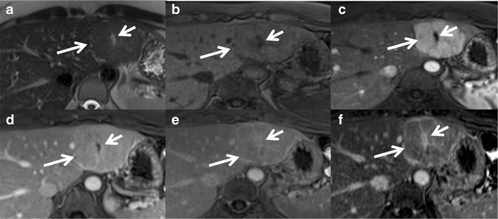
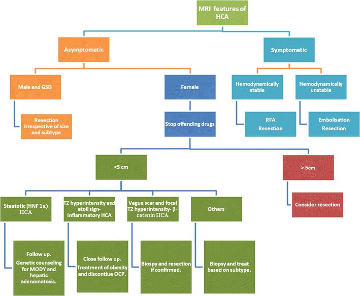
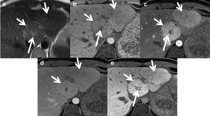
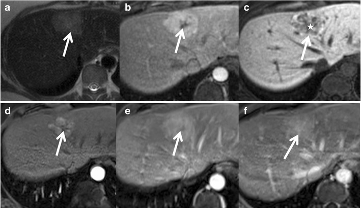
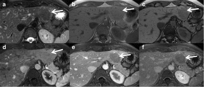
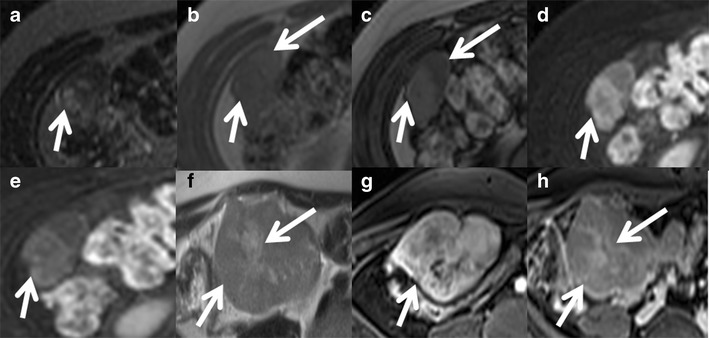
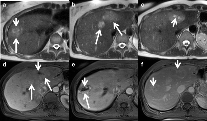
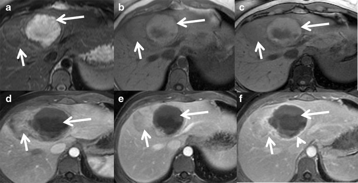
Focal nodular hyperplasia (FNH) and hepatocellular adenomas (HCAs) constitute benign hepatic neoplasms in adults. HCAs are monoclonal neoplasms characterised by an increased predilection to haemorrhage and also malignant transformation. On the other hand, FNH is a polyclonal tumour-like lesion that occurs in response to increased perfusion and has an uneventful clinical course. Recent advances in molecular genetics and genotype-phenotype correlation in these hepatocellular neoplasms have enabled a new classification system. FNHs are classified into the typical and atypical types based on histomorphological and imaging features. HCAs have been categorised into four subtypes: (1) HCAs with HNF-1α mutations are diffusely steatotic, do not undergo malignant transformation, and are associated with familial diabetes or adenomatosis. (2) Inflammatory HCAs are hypervascular with marked peliosis and a tendency to bleed. They are associated with obesity, alcohol and hepatic steatosis. (3) HCAs with β-catenin mutations are associated with male hormone administration and glycogen storage disease, frequently undergo malignant transformation and may simulate hepatocellular carcinoma on imaging. (4) The final type is unclassified HCAs. Each of these except the unclassified subtype has a few distinct imaging features, often enabling reasonably accurate diagnosis. Biopsy with immunohistochemical analysis is helpful in difficult cases and has strong implications for patient management.

TEACHING POINTS

• FNHs are benign polyclonal neoplasms with no risk of haemorrhage or malignancy. • HCAs are benign monoclonal neoplasms classified into four subtypes based on immunohistochemistry. • Inflammatory HCAs show an atoll sign with a risk of bleeding and malignant transformation. • HNF-1α HCAs are steatotic HCAs with minimal complications and the best prognosis. • β-Catenin HCA shows variable MRI features and a high risk of malignancy.

摘要

未标注

局灶性结节性增生(FNH)和肝细胞腺瘤(HCA)是成人肝脏的良性肿瘤。HCA是单克隆肿瘤,其特点是出血倾向增加,也有恶变倾向。另一方面,FNH是一种多克隆肿瘤样病变,由灌注增加引起,临床过程平稳。这些肝细胞肿瘤在分子遗传学和基因型-表型相关性方面的最新进展促成了一种新的分类系统。FNH根据组织形态学和影像学特征分为典型和非典型类型。HCA已被分为四种亚型:(1)具有HNF-1α突变的HCA弥漫性脂肪变性,不会发生恶变,与家族性糖尿病或腺瘤病相关。(2)炎症性HCA血管丰富,有明显的血囊肿形成且有出血倾向。它们与肥胖、酒精和肝脂肪变性有关。(3)具有β-连环蛋白突变的HCA与雄激素给药和糖原贮积病有关,经常发生恶变,在影像学上可能类似肝细胞癌。(4)最后一种类型是未分类的HCA。除未分类亚型外,每种亚型都有一些独特的影像学特征,通常能够进行合理准确的诊断。免疫组织化学分析活检对疑难病例有帮助,对患者管理有重要意义。

教学要点

•FNH是良性多克隆肿瘤,无出血或恶变风险。•HCA是良性单克隆肿瘤,根据免疫组织化学分为四种亚型。•炎症性HCA显示环礁征,有出血和恶变风险。•HNF-1α HCA是脂肪变性的HCA,并发症最少,预后最佳。•β-连环蛋白HCA显示MRI特征多样,恶变风险高。

https://cdn.ncbi.nlm.nih.gov/pmc/blobs/57f7/4444792/36321be16f54/13244_2015_399_Fig13_HTML.jpg
https://cdn.ncbi.nlm.nih.gov/pmc/blobs/57f7/4444792/8898fdf4f1ea/13244_2015_399_Fig1_HTML.jpg
https://cdn.ncbi.nlm.nih.gov/pmc/blobs/57f7/4444792/a40fc358fc84/13244_2015_399_Fig2_HTML.jpg
https://cdn.ncbi.nlm.nih.gov/pmc/blobs/57f7/4444792/e5178f9be20e/13244_2015_399_Fig3_HTML.jpg
https://cdn.ncbi.nlm.nih.gov/pmc/blobs/57f7/4444792/9e0e126cf77b/13244_2015_399_Fig4_HTML.jpg
https://cdn.ncbi.nlm.nih.gov/pmc/blobs/57f7/4444792/c69d3344bdd8/13244_2015_399_Fig5_HTML.jpg
https://cdn.ncbi.nlm.nih.gov/pmc/blobs/57f7/4444792/c35253a4fad4/13244_2015_399_Fig6_HTML.jpg
https://cdn.ncbi.nlm.nih.gov/pmc/blobs/57f7/4444792/05690b58cb2a/13244_2015_399_Fig7_HTML.jpg
https://cdn.ncbi.nlm.nih.gov/pmc/blobs/57f7/4444792/71226c432b9c/13244_2015_399_Fig8_HTML.jpg
https://cdn.ncbi.nlm.nih.gov/pmc/blobs/57f7/4444792/140675322ebd/13244_2015_399_Fig9_HTML.jpg
https://cdn.ncbi.nlm.nih.gov/pmc/blobs/57f7/4444792/5eed6ed478a8/13244_2015_399_Fig10_HTML.jpg
https://cdn.ncbi.nlm.nih.gov/pmc/blobs/57f7/4444792/74107fdcea39/13244_2015_399_Fig11_HTML.jpg
https://cdn.ncbi.nlm.nih.gov/pmc/blobs/57f7/4444792/dc20a7618daa/13244_2015_399_Fig12_HTML.jpg
https://cdn.ncbi.nlm.nih.gov/pmc/blobs/57f7/4444792/36321be16f54/13244_2015_399_Fig13_HTML.jpg

相似文献

1
Current updates on the molecular genetics and magnetic resonance imaging of focal nodular hyperplasia and hepatocellular adenoma.局灶性结节性增生和肝细胞腺瘤的分子遗传学与磁共振成像的最新进展
Insights Imaging. 2015 Jun;6(3):347-62. doi: 10.1007/s13244-015-0399-8. Epub 2015 Mar 20.
2
Hepatocyte Nuclear Factor 1α-Mutated Hepatocellular Adenomas: An Atypical Presentation.肝细胞核因子1α突变型肝细胞腺瘤:一种非典型表现。
Case Rep Gastroenterol. 2017 Oct 30;11(3):637-642. doi: 10.1159/000480376. eCollection 2017 Sep-Dec.
3
Hepatocellular Adenoma Subtypes Based on 2017 Classification System: Exploratory Study of Gadoxetate Disodium-Enhanced MRI Features With Proposal of a Diagnostic Algorithm.基于 2017 分类系统的肝细胞腺瘤亚型:钆塞酸二钠增强 MRI 特征的探索性研究及诊断算法的提出。
AJR Am J Roentgenol. 2023 Apr;220(4):539-550. doi: 10.2214/AJR.22.28233. Epub 2022 Sep 28.
4
Molecular Classification of Hepatocellular Adenoma Associates With Risk Factors, Bleeding, and Malignant Transformation.肝细胞腺瘤的分子分类与危险因素、出血和恶性转化相关。
Gastroenterology. 2017 Mar;152(4):880-894.e6. doi: 10.1053/j.gastro.2016.11.042. Epub 2016 Dec 7.
5
Recent advances in cytogenetics and molecular biology of adult hepatocellular tumors: implications for imaging and management.成人肝细胞肿瘤细胞遗传学和分子生物学的最新进展:对影像学和治疗的影响。
Radiology. 2011 Mar;258(3):673-93. doi: 10.1148/radiol.10100376.
6
New MRI features improve subtype classification of hepatocellular adenoma.新的 MRI 特征可改善肝细胞腺瘤的亚型分类。
Eur Radiol. 2019 May;29(5):2436-2447. doi: 10.1007/s00330-018-5784-5. Epub 2018 Dec 6.
7
Hepatocellular adenomas: magnetic resonance imaging features as a function of molecular pathological classification.肝细胞腺瘤:磁共振成像特征与分子病理分类的关系
Hepatology. 2008 Sep;48(3):808-18. doi: 10.1002/hep.22417.
8
[Focal nodular hyperplasia and hepatocellular adenoma].[局灶性结节性增生与肝细胞腺瘤]
Radiologe. 2015 Jan;55(1):18-26. doi: 10.1007/s00117-014-2704-9.
9
Quantitative correlation between uptake of Gd-BOPTA on hepatobiliary phase and tumor molecular features in patients with benign hepatocellular lesions.良性肝细胞病变患者肝胆期摄取 Gd-BOPTA 与肿瘤分子特征的定量相关性。
Eur Radiol. 2018 Oct;28(10):4243-4253. doi: 10.1007/s00330-018-5438-7. Epub 2018 May 2.
10
A Shifting Paradigm in Diagnosis and Management of Hepatic Adenoma.肝腺瘤的诊断与治疗范式转变
Ann Surg Oncol. 2020 Sep;27(9):3330-3338. doi: 10.1245/s10434-020-08580-w. Epub 2020 Jun 15.

引用本文的文献

1
A T2 weighted imaging-based radiomics nomogram for the classification of hepatic blood-rich lesions: hepatocellular carcinoma and benign liver lesions.基于T2加权成像的放射组学列线图用于肝血供丰富病变的分类:肝细胞癌和良性肝病变
Discov Oncol. 2025 Jul 1;16(1):1223. doi: 10.1007/s12672-025-02868-7.
2
Qualitative and quantitative assessment of gadoxetic acid MRI in distinguishing atypical focal nodular hyperplasia from hepatocellular adenoma subtypes.钆塞酸MRI在鉴别非典型性局灶性结节性增生与肝细胞腺瘤亚型中的定性和定量评估
Eur Radiol. 2025 May 14. doi: 10.1007/s00330-025-11679-x.
3
Correlation between magnetic resonance imaging (MRI) features and lesion size in focal nodular hyperplasia of the liver.

本文引用的文献

1
Inflammatory hepatocellular adenomas can mimic focal nodular hyperplasia on gadoxetic acid-enhanced MRI.炎症性肝细胞腺瘤在钆塞酸增强磁共振成像(MRI)上可模仿局灶性结节性增生。
AJR Am J Roentgenol. 2014 Oct;203(4):W408-14. doi: 10.2214/AJR.13.12251. Epub 2014 Jul 23.
2
Risk factors for bleeding in hepatocellular adenoma.肝细胞腺瘤出血的风险因素。
Br J Surg. 2014 Jun;101(7):847-55. doi: 10.1002/bjs.9493. Epub 2014 Apr 24.
3
MRI features of inflammatory hepatocellular adenomas on hepatocyte phase imaging with liver-specific contrast agents.
肝脏局灶性结节性增生的磁共振成像(MRI)特征与病变大小之间的相关性
Quant Imaging Med Surg. 2024 Dec 5;14(12):8758-8770. doi: 10.21037/qims-24-836. Epub 2024 Nov 13.
4
Gd-EOB-DTPA enhanced MRI nomogram model to differentiate hepatocellular carcinoma and focal nodular hyperplasia both showing iso- or hyperintensity in the hepatobiliary phase.钆塞酸二钠增强 MRI 列线图模型鉴别肝胆期呈等或高信号的肝细胞肝癌和局灶性结节增生。
BMC Med Imaging. 2024 Aug 12;24(1):211. doi: 10.1186/s12880-024-01382-6.
5
Giant focal nodular hyperplasia with a background of hepatic steatosis in a 14-year-old boy.一名14岁男孩患有伴有肝脂肪变性背景的巨大局灶性结节性增生。
J Surg Case Rep. 2022 May 27;2022(5):rjac238. doi: 10.1093/jscr/rjac238. eCollection 2022 May.
6
A Scoping Review of the Classification, Diagnosis, and Management of Hepatic Adenomas.肝腺瘤分类、诊断及管理的范围综述
J Gastrointest Surg. 2022 Apr;26(4):965-978. doi: 10.1007/s11605-022-05246-8. Epub 2022 Jan 26.
7
Ultrasound findings in peliosis hepatis.肝紫癜病的超声表现
Ultrasonography. 2021 Oct;40(4):546-554. doi: 10.14366/usg.20162. Epub 2021 Feb 22.
8
Overview of spontaneous intraabdominal tumor hemorrhage: etiologies, imaging findings, and management.自发性腹腔内肿瘤出血概述:病因、影像学表现及处理。
Abdom Radiol (NY). 2021 Feb;46(2):427-440. doi: 10.1007/s00261-020-02663-8. Epub 2020 Jul 20.
9
Patterns of enhancement in the hepatobiliary phase of gadoxetic acid-enhanced MRI.钆塞酸增强MRI肝胆期的强化模式。
Br J Radiol. 2020 Aug;93(1112):20190989. doi: 10.1259/bjr.20190989. Epub 2020 Jun 1.
10
A CT-based radiomics nomogram for differentiation of focal nodular hyperplasia from hepatocellular carcinoma in the non-cirrhotic liver.基于 CT 的放射组学列线图用于区分非肝硬化肝脏中的局灶性结节性增生与肝细胞癌。
Cancer Imaging. 2020 Feb 24;20(1):20. doi: 10.1186/s40644-020-00297-z.
使用肝脏特异性对比剂进行肝细胞期成像时炎症性肝细胞腺瘤的MRI特征
J Magn Reson Imaging. 2014 May;39(5):1259-64. doi: 10.1002/jmri.24281. Epub 2013 Jul 29.
4
Focal Nodular Hyperplasia and Hepatocellular Adenoma around the World Viewed through the Scope of the Immunopathological Classification.通过免疫病理分类视角看世界各地的局灶性结节性增生和肝细胞腺瘤
Int J Hepatol. 2013;2013:268625. doi: 10.1155/2013/268625. Epub 2013 Apr 14.
5
Immunohistochemical markers on needle biopsies are helpful for the diagnosis of focal nodular hyperplasia and hepatocellular adenoma subtypes.针吸活检的免疫组化标志物有助于局灶性结节性增生和肝细胞腺瘤亚型的诊断。
Am J Surg Pathol. 2012 Nov;36(11):1691-9. doi: 10.1097/PAS.0b013e3182653ece.
6
Diagnostic accuracy of MRI in differentiating hepatocellular adenoma from focal nodular hyperplasia: prospective study of the additional value of gadoxetate disodium.MRI 对肝细胞腺瘤与局灶性结节增生的诊断准确性:钆塞酸二钠的附加价值的前瞻性研究。
AJR Am J Roentgenol. 2012 Jul;199(1):26-34. doi: 10.2214/AJR.11.7750.
7
Focal nodular hyperplasia: hepatobiliary enhancement patterns on gadoxetic-acid contrast-enhanced MRI.局灶性结节性增生:钆塞酸二钠增强MRI上的肝胆强化模式
Abdom Imaging. 2013 Jun;38(3):490-501. doi: 10.1007/s00261-012-9916-0.
8
Focal hepatic lesions in Gd-EOB-DTPA enhanced MRI: the atlas.钆塞酸二钠增强 MRI 中的局灶性肝脏病变:图谱。
Insights Imaging. 2012 Oct;3(5):451-74. doi: 10.1007/s13244-012-0179-7. Epub 2012 Jun 15.
9
Systematic review of haemorrhage and rupture of hepatocellular adenomas.系统综述肝细胞腺瘤的出血和破裂。
Br J Surg. 2012 Jul;99(7):911-6. doi: 10.1002/bjs.8762. Epub 2012 May 22.
10
Characteristics and distinguishing features of hepatocellular adenoma and focal nodular hyperplasia on gadoxetate disodium-enhanced MRI.钆塞酸二钠增强 MRI 诊断肝细胞腺瘤和局灶性结节增生的特征和鉴别特征。
AJR Am J Roentgenol. 2012 Jan;198(1):115-23. doi: 10.2214/AJR.11.6836.