Cortes Daniel H, Suydam Stephen M, Silbernagel Karin Grävare, Buchanan Thomas S, Elliott Dawn M
Biomedical Engineering Program, University of Delaware, Newark, Delaware, USA.
Mechanical Engineering Department, University of Delaware, Newark, Delaware, USA.
Ultrasound Med Biol. 2015 Jun;41(6):1518-29. doi: 10.1016/j.ultrasmedbio.2015.02.001. Epub 2015 Mar 19.
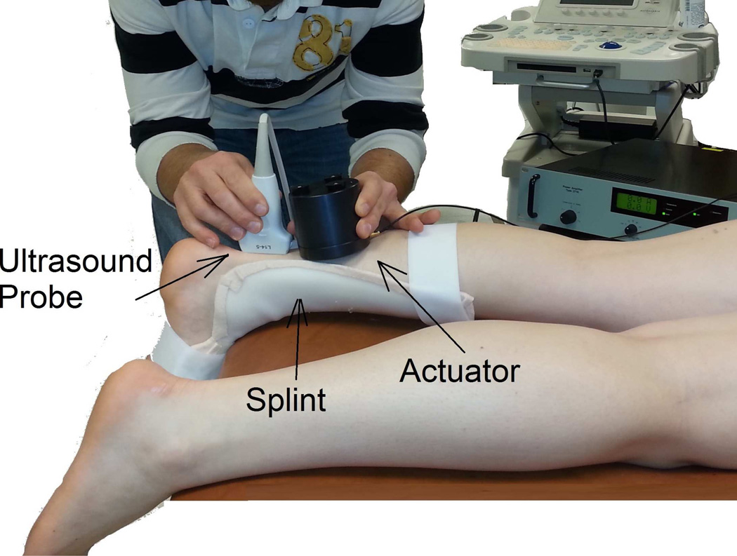
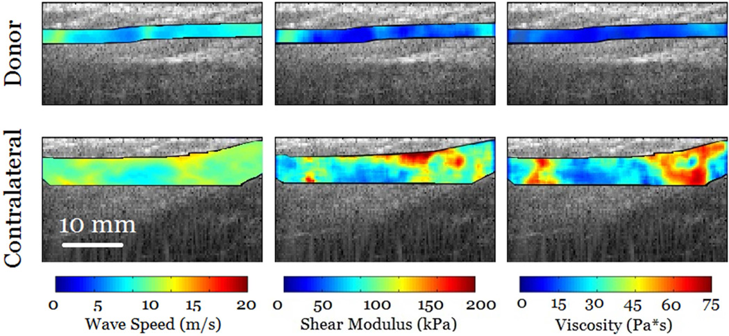
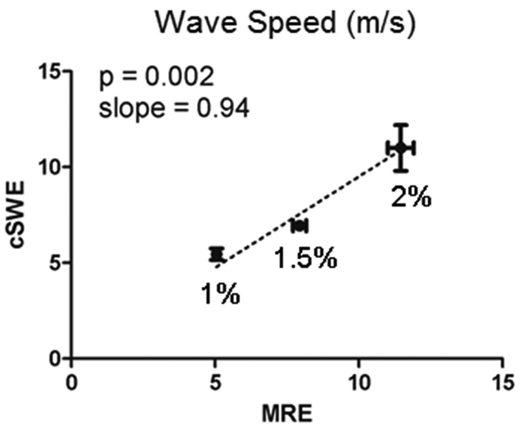
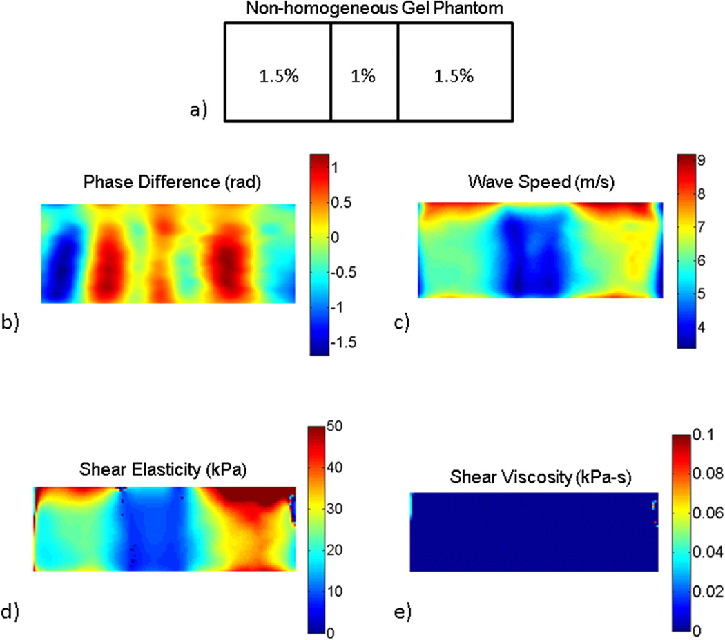
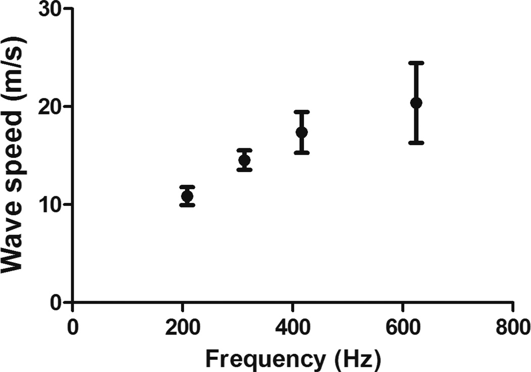
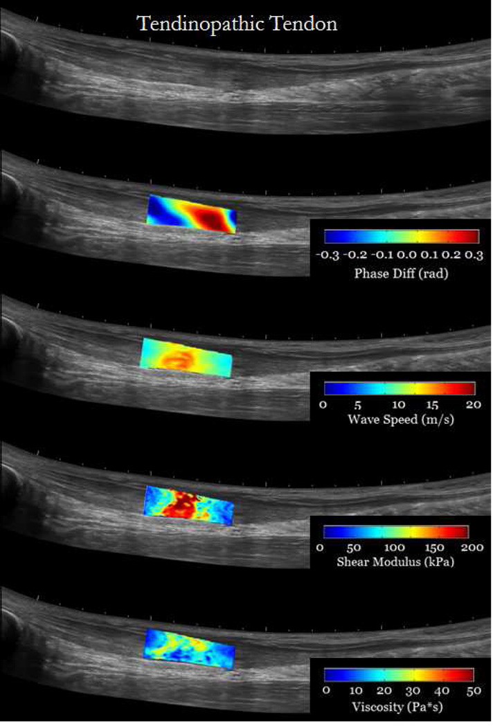
Viscoelastic mechanical properties are frequently altered after tendon injuries and during recovery. Therefore, non-invasive measurements of shear viscoelastic properties may help evaluate tendon recovery and compare the effectiveness of different therapies. The objectives of this study were to describe an elastography method for measuring localized viscoelastic properties of tendons and to discuss the initial results in healthy and injured human Achilles and semitendinosus tendons. The technique used an external actuator to generate the shear waves in the tendon at different frequencies and plane wave imaging to measure shear wave displacements. For each of the excitation frequencies, maps of direction-specific wave speeds were calculated using local frequency estimation. Maps of viscoelastic properties were obtained using a pixel-wise curve fit of wave speed and frequency. The method was validated by comparing measurements of wave speed in agarose gels with those obtained using magnetic resonance elastography. Measurements in human healthy Achilles tendons revealed a pronounced increase in wave speed as a function of frequency, which highlights the importance of tendon viscoelasticity. Additionally, the viscoelastic properties of the Achilles tendon were larger than those reported for other tissues. Measurements in a tendinopathic Achilles tendon indicated that it is feasible to quantify local viscoelastic properties. Similarly, measurement in the semitendinosus tendon revealed substantial differences in viscoelastic properties between the healthy and contralateral tendons. Consequently, this technique has the potential to evaluate localized changes in tendon viscoelastic properties caused by injury and during recovery in a clinical setting.
肌腱损伤后及恢复过程中,其粘弹性力学特性常发生改变。因此,对剪切粘弹性特性进行非侵入性测量可能有助于评估肌腱恢复情况,并比较不同治疗方法的效果。本研究的目的是描述一种用于测量肌腱局部粘弹性特性的弹性成像方法,并讨论在健康及受伤的人类跟腱和半腱肌肌腱中的初步结果。该技术使用外部致动器在不同频率下在肌腱中产生剪切波,并利用平面波成像测量剪切波位移。对于每个激发频率,使用局部频率估计计算特定方向波速的图谱。通过对波速和频率进行逐像素曲线拟合获得粘弹性特性图谱。通过将琼脂糖凝胶中的波速测量结果与磁共振弹性成像获得的结果进行比较,对该方法进行了验证。在人类健康跟腱中的测量结果显示,波速随频率显著增加,这突出了肌腱粘弹性的重要性。此外,跟腱的粘弹性特性大于其他组织的报道值。在患有肌腱病的跟腱中的测量表明,量化局部粘弹性特性是可行的。同样,在半腱肌肌腱中的测量显示,健康肌腱与对侧肌腱的粘弹性特性存在显著差异。因此,该技术有潜力在临床环境中评估由损伤及恢复过程引起的肌腱粘弹性特性的局部变化。