Richter Torsten, Bergmann Ralf, Musch Guido, Pietzsch Jens, Koch Thea
Department of Anesthesia and Intensive Care, Carl Gustav Carus University Hospital, Technische Universität Dresden, Dresden, Germany.
Department of Radiopharmaceutical and Chemical Biology, Institute of Radiopharmaceutical Cancer Research, Helmholtz-Zentrum Dresden- Rossendorf, Dresden, Germany.
BMC Anesthesiol. 2015 Mar 18;15:36. doi: 10.1186/s12871-015-0013-0. eCollection 2015.
Aspiration-induced lung injury can decrease gas exchange and increase mortality. Acute lung injury following acid aspiration is characterized by elevated pulmonary blood flow (PBF) in damaged lung areas in the early inflammation stage. Knowledge of PBF patterns after acid aspiration is important for targeting intravenous treatments. We examined PBF in an experimental model at a later stage (2 hours after injury).
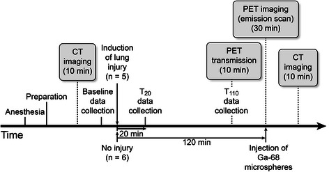
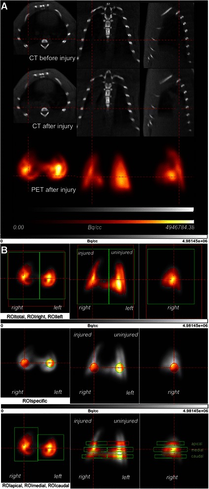
Anesthetized Wistar-Unilever rats (n = 5) underwent unilateral endobronchial instillation of hydrochloric acid. The PBF distribution was compared between injured and uninjured sides and with that of untreated control animals (n = 6). Changes in lung density after injury were measured using computed tomography (CT). Regional PBF distribution was determined quantitatively in vivo 2 hours after acid instillation by measuring the concentration of [(68)Ga]-radiolabeled microspheres using positron emission tomography.
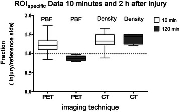
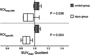
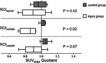
CT scans revealed increased lung density in areas of acid aspiration. Lung injury was accompanied by impaired gas exchange. Acid aspiration decreased the arterial pressure of oxygen from 157 mmHg [139;165] to 74 mmHg [67;86] at 20 minutes and tended toward restoration to 109 mmHg [69;114] at 110 minutes (P < 0.001). The PBF ratio of the middle region of the injured versus uninjured lungs of the aspiration group (0.86 [0.7;0.9], median [25%;75%]) was significantly lower than the PBF ratio in the left versus right lung of the control group (1.02 [1.0;1.05]; P = 0.016).
The PBF pattern 2 hours after aspiration-induced lung injury showed a redistribution of PBF away from injured regions that was likely responsible for the partial recovery from hypoxemia over time. Treatments given intravenously 2 hours after acid-induced lung injury may not preferentially reach the injured lung regions, contrary to what occurs during the first hour of inflammation. Please see related article: http://dx.doi.org/10.1186/s12871-015-0014-z.
误吸所致肺损伤可降低气体交换并增加死亡率。酸误吸后的急性肺损伤在炎症早期的特征是受损肺区肺血流量(PBF)升高。了解酸误吸后的PBF模式对于靶向静脉治疗很重要。我们在损伤后期(损伤后2小时)的实验模型中检测了PBF。
对麻醉的Wistar-Unilever大鼠(n = 5)进行单侧支气管内滴注盐酸。比较损伤侧和未损伤侧的PBF分布,并与未治疗的对照动物(n = 6)进行比较。使用计算机断层扫描(CT)测量损伤后肺密度的变化。通过正电子发射断层扫描测量[(68)Ga]放射性标记微球的浓度,在酸滴注后2小时在体内定量测定区域PBF分布。
CT扫描显示酸误吸区域肺密度增加。肺损伤伴有气体交换受损。酸误吸使氧分压在20分钟时从157 mmHg [139;165]降至74 mmHg [67;86],并在110分钟时倾向于恢复至109 mmHg [69;114](P < 0.001)。误吸组损伤肺与未损伤肺中间区域的PBF比值(0.86 [0.7;0.9],中位数[25%;75%])显著低于对照组左肺与右肺的PBF比值(1.02 [1.0;1.05];P = 0.016)。
误吸所致肺损伤后2小时的PBF模式显示PBF从损伤区域重新分布,这可能是低氧血症随时间部分恢复的原因。与炎症的第一小时情况相反,酸诱导肺损伤后2小时静脉给予的治疗可能不会优先到达损伤肺区域。请参阅相关文章:http://dx.doi.org/10.1186/s12871-015-0014-z。