Lapa Constantin, Linsenmann Thomas, Lückerath Katharina, Samnick Samuel, Herrmann Ken, Stoffer Carolin, Ernestus Ralf-Ingo, Buck Andreas K, Löhr Mario, Monoranu Camelia-Maria
Department of Nuclear Medicine, University Hospital Würzburg, Würzburg, Germany.
Department of Neurosurgery, University Hospital Würzburg, Würzburg, Germany.
PLoS One. 2015 Mar 25;10(3):e0122269. doi: 10.1371/journal.pone.0122269. eCollection 2015.
Glioblastoma multiforme (GBM) is the most common primary brain tumor in adults. Tumor-associated macrophages (TAM) have been shown to promote malignant growth and to correlate with poor prognosis. [1,4,7,10-tetraazacyclododecane-NN',N″,N'″-tetraacetic acid]-d-Phe1,Tyr3-octreotate (DOTATATE) labeled with Gallium-68 selectively binds to somatostatin receptor 2A (SSTR2A) which is specifically expressed and up-regulated in activated macrophages. On the other hand, the role of SSTR2A expression on the cell surface of glioma cells has not been fully elucidated yet. The aim of this study was to non-invasively assess SSTR2A expression of both glioma cells as well as macrophages in GBM.
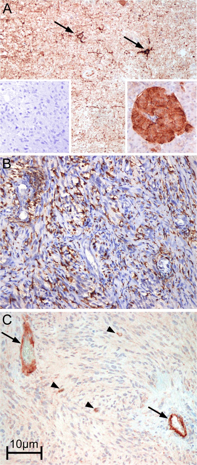
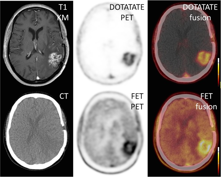
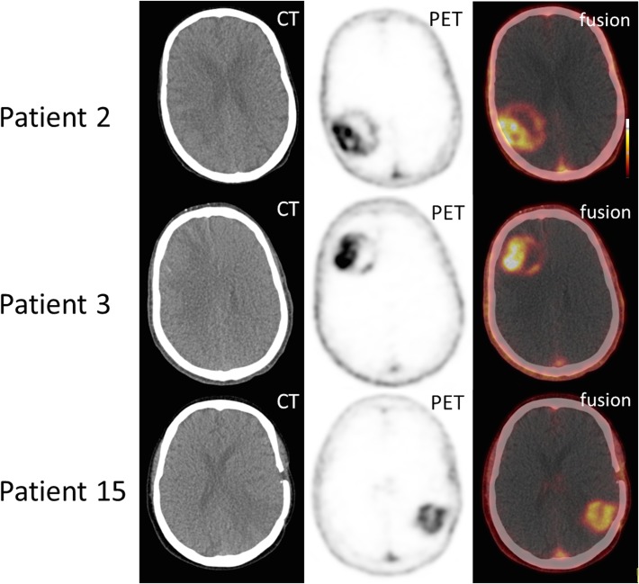
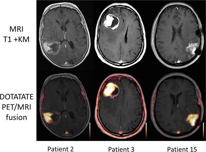
15 samples of patient-derived GBM were stained immunohistochemically for macrophage infiltration (CD68), proliferative activity (Ki67) as well as expression of SSTR2A. Anti-CD45 staining was performed to distinguish between resident microglia and tumor-infiltrating macrophages. In a subcohort, positron emission tomography (PET) imaging using 68Ga-DOTATATE was performed and the semiquantitatively evaluated tracer uptake was compared to the results of immunohistochemistry.
The amount of microglia/macrophages ranged from <10% to >50% in the tumor samples with the vast majority being resident microglial cells. A strong SSTR2A immunostaining was observed in endothelial cells of proliferating vessels, in neurons and neuropile. Only faint immunostaining was identified on isolated microglial and tumor cells. Somatostatin receptor imaging revealed areas of increased tracer accumulation in every patient. However, retention of the tracer did not correlate with immunohistochemical staining patterns.
SSTR2A seems not to be overexpressed in GBM samples tested, neither on the cell surface of resident microglia or infiltrating macrophages, nor on the surface of tumor cells. These data suggest that somatostatin receptor directed imaging and treatment strategies are less promising in GBM.
多形性胶质母细胞瘤(GBM)是成人中最常见的原发性脑肿瘤。肿瘤相关巨噬细胞(TAM)已被证明可促进恶性生长并与预后不良相关。[1,4,7,10-四氮杂环十二烷-NN′,N″,N′″-四乙酸]-d-苯丙氨酸1,酪氨酸3-奥曲肽(DOTATATE)用镓-68标记后可选择性结合生长抑素受体2A(SSTR2A),该受体在活化巨噬细胞中特异性表达并上调。另一方面,SSTR2A在胶质瘤细胞表面表达的作用尚未完全阐明。本研究的目的是无创评估GBM中胶质瘤细胞以及巨噬细胞的SSTR2A表达。
对15例患者来源的GBM样本进行免疫组织化学染色,检测巨噬细胞浸润(CD68)、增殖活性(Ki67)以及SSTR2A的表达。进行抗CD45染色以区分常驻小胶质细胞和肿瘤浸润巨噬细胞。在一个亚组中,使用68Ga-DOTATATE进行正电子发射断层扫描(PET)成像,并将半定量评估的示踪剂摄取与免疫组织化学结果进行比较。
肿瘤样本中微胶质细胞/巨噬细胞的数量范围从<10%到>50%,绝大多数是常驻微胶质细胞。在增殖血管的内皮细胞、神经元和神经纤维中观察到强烈的SSTR2A免疫染色。在分离的微胶质细胞和肿瘤细胞上仅发现微弱的免疫染色。生长抑素受体成像显示每个患者中示踪剂积累增加的区域。然而,示踪剂的滞留与免疫组织化学染色模式无关。
在测试的GBM样本中,SSTR2A似乎未过度表达,无论是在常驻微胶质细胞或浸润巨噬细胞的细胞表面,还是在肿瘤细胞表面。这些数据表明,生长抑素受体导向的成像和治疗策略在GBM中前景不太乐观。