• 文献检索
  • 文档翻译
  • 深度研究
  • 学术资讯
  • Suppr Zotero 插件Zotero 插件
  • 邀请有礼
  • 套餐&价格
  • 历史记录
应用&插件
Suppr Zotero 插件Zotero 插件浏览器插件Mac 客户端Windows 客户端微信小程序
定价
高级版会员购买积分包购买API积分包
服务
文献检索文档翻译深度研究API 文档MCP 服务
关于我们
关于 Suppr公司介绍联系我们用户协议隐私条款
关注我们

Suppr 超能文献

核心技术专利:CN118964589B侵权必究
粤ICP备2023148730 号-1Suppr @ 2026

文献检索

告别复杂PubMed语法,用中文像聊天一样搜索,搜遍4000万医学文献。AI智能推荐,让科研检索更轻松。

立即免费搜索

文件翻译

保留排版,准确专业,支持PDF/Word/PPT等文件格式,支持 12+语言互译。

免费翻译文档

深度研究

AI帮你快速写综述,25分钟生成高质量综述,智能提取关键信息,辅助科研写作。

立即免费体验

乳腺癌脑微转移的在线相衬和基于光栅的相衬同步辐射成像研究

In-line phase-contrast and grating-based phase-contrast synchrotron imaging study of brain micrometastasis of breast cancer.

作者信息

Huang Sheng, Kou Binquan, Chi Yayun, Xi Yan, Cao Yixin, Cui Wenli, Hu Xin, Shao Zhimin, Guo Han, Fu Yanan, Xiao Tiqiao, Sun Jianqi, Zhao Jun, Wang Yujie, Wu Jiong

机构信息

Department of Breast Surgery, Breast Cancer Institute, Shanghai Cancer Center, Department of Oncology, Shanghai Medical College, Fudan University, Shanghai, People's Republic of China.

Department of Physics and Astronomy, Shanghai Jiao Tong University, Shanghai, People's Republic of China.

出版信息

Sci Rep. 2015 Mar 30;5:9418. doi: 10.1038/srep09418.

DOI:10.1038/srep09418
PMID:25818989
原文链接:https://pmc.ncbi.nlm.nih.gov/articles/PMC4377630/
Abstract

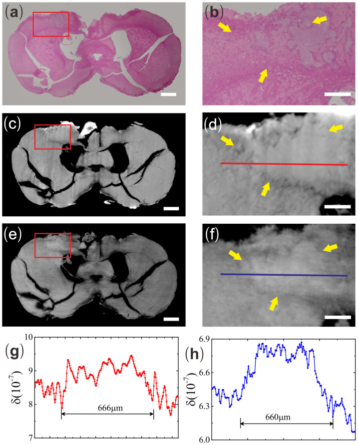
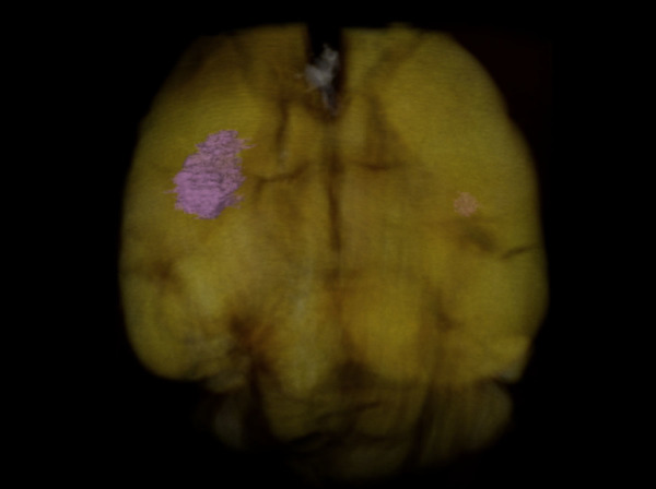
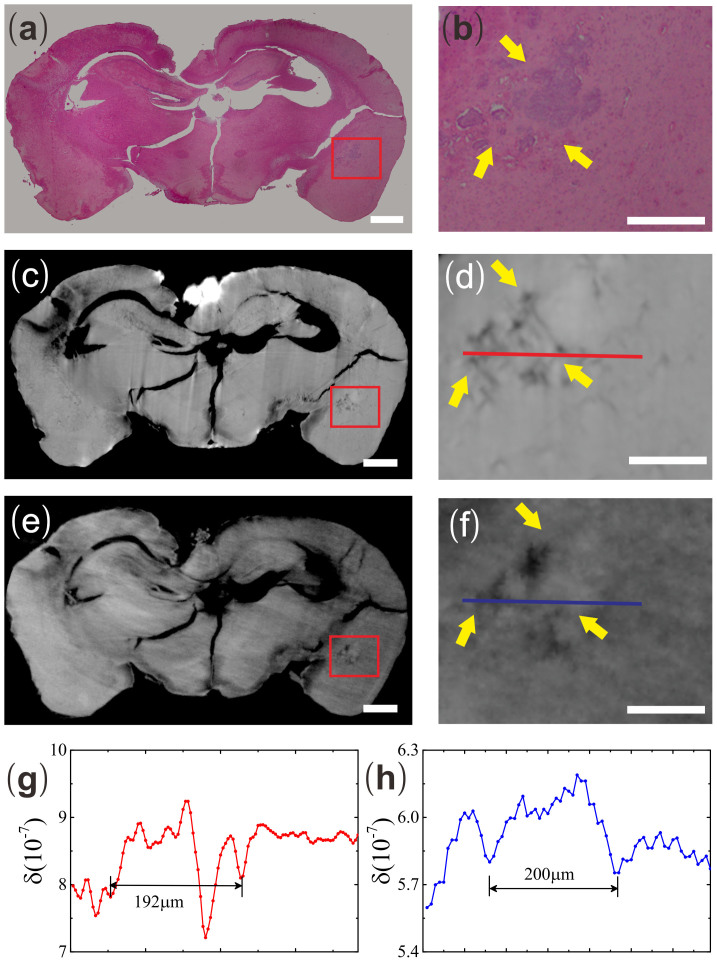
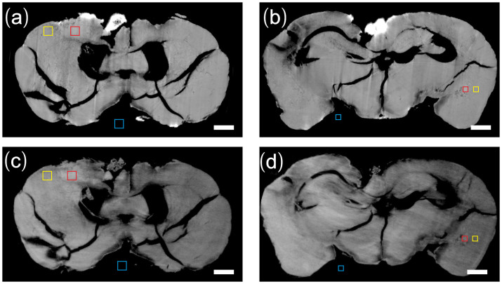
Current bio-medical imaging researches aim to detect brain micrometastasis in early stage for its increasing incidence and high mortality rates. Synchrotron phase-contrast imaging techniques, such as in-line phase-contrast (IPC) and grating-based phase-contrast (GPC) imaging, could provide a high spatial and density imaging study of biological specimens' 3D structures. In this study, we demonstrated the detection efficiencies of these two imaging tools on breast cancer micrometastasis in an ex vivo mouse brain. We found that both IPC and GPC can differentiate abnormal brain structures induced by micrometastasis from the surrounding normal tissues. We also found that GPC was more sensitive in detecting the small metastasis as compared to IPC.

摘要

当前生物医学成像研究旨在早期检测脑微转移,因为其发病率不断上升且死亡率很高。同步辐射相衬成像技术,如同轴相衬(IPC)和基于光栅的相衬(GPC)成像,可以对生物标本的三维结构进行高空间分辨率和密度的成像研究。在本研究中,我们展示了这两种成像工具在体外小鼠脑内对乳腺癌微转移的检测效率。我们发现IPC和GPC都能将微转移诱导的异常脑结构与周围正常组织区分开来。我们还发现,与IPC相比,GPC在检测小转移灶方面更敏感。

https://cdn.ncbi.nlm.nih.gov/pmc/blobs/d65a/4377630/25f0f9bdc78e/srep09418-f4.jpg
https://cdn.ncbi.nlm.nih.gov/pmc/blobs/d65a/4377630/c482cb381c47/srep09418-f1.jpg
https://cdn.ncbi.nlm.nih.gov/pmc/blobs/d65a/4377630/e62f4613de5c/srep09418-f2.jpg
https://cdn.ncbi.nlm.nih.gov/pmc/blobs/d65a/4377630/d51e711beb9d/srep09418-f3.jpg
https://cdn.ncbi.nlm.nih.gov/pmc/blobs/d65a/4377630/25f0f9bdc78e/srep09418-f4.jpg

相似文献

1
In-line phase-contrast and grating-based phase-contrast synchrotron imaging study of brain micrometastasis of breast cancer.乳腺癌脑微转移的在线相衬和基于光栅的相衬同步辐射成像研究
Sci Rep. 2015 Mar 30;5:9418. doi: 10.1038/srep09418.
2
Lung cancer and angiogenesis imaging using synchrotron radiation.利用同步辐射进行肺癌和血管生成成像。
Phys Med Biol. 2010 Apr 21;55(8):2399-409. doi: 10.1088/0031-9155/55/8/017. Epub 2010 Apr 1.
3
Label-free FTIR spectroscopy detects and visualizes the early stage of pulmonary micrometastasis seeded from breast carcinoma.无标记傅里叶变换红外光谱检测并可视化源自乳腺癌的肺微转移的早期阶段。
Biochim Biophys Acta Mol Basis Dis. 2018 Nov;1864(11):3574-3584. doi: 10.1016/j.bbadis.2018.08.022. Epub 2018 Aug 18.
4
Phase contrast imaging of breast tumours with synchrotron radiation.利用同步辐射对乳腺肿瘤进行相衬成像。
Appl Radiat Isot. 2009 Jun;67(6):1033-41. doi: 10.1016/j.apradiso.2009.01.075. Epub 2009 Feb 26.
5
Validation of a technique using microbubbles and contrast enhanced ultrasound (CEUS) to biopsy sentinel lymph nodes (SLN) in pre-operative breast cancer patients with a normal grey-scale axillary ultrasound.验证一种使用微泡和对比增强超声(CEUS)对术前腋窝超声正常的乳腺癌患者进行前哨淋巴结(SLN)活检的技术。
Eur J Surg Oncol. 2013 Jul;39(7):760-5. doi: 10.1016/j.ejso.2013.03.026. Epub 2013 Apr 28.
6
Quantitative breast tissue characterization using grating-based x-ray phase-contrast imaging.使用基于光栅的X射线相衬成像进行乳腺组织定量表征。
Phys Med Biol. 2014 Apr 7;59(7):1557-71. doi: 10.1088/0031-9155/59/7/1557. Epub 2014 Mar 10.
7
Evaluation of phase-contrast CT of breast tissue at conventional X-ray sources - presentation of selected findings.常规 X 射线源下乳腺组织的相位对比 CT 评估 - 部分发现的呈现。
Z Med Phys. 2013 Sep;23(3):212-21. doi: 10.1016/j.zemedi.2013.02.005. Epub 2013 Mar 11.
8
Diffraction enhanced imaging contrast mechanisms in breast cancer specimens.乳腺癌标本中的衍射增强成像对比机制。
Med Phys. 2002 Oct;29(10):2216-21. doi: 10.1118/1.1507782.
9
X-ray phase-contrast tomosynthesis for improved breast tissue discrimination.X 射线相衬断层合成术提高乳腺组织的分辨能力。
Eur J Radiol. 2014 Mar;83(3):531-6. doi: 10.1016/j.ejrad.2013.12.005. Epub 2013 Dec 12.
10
Advanced contrast modalities for X-ray radiology: Phase-contrast and dark-field imaging using a grating interferometer.X 射线放射学的高级对比模式:使用光栅干涉仪的相位对比和暗场成像。
Z Med Phys. 2010;20(1):7-16. doi: 10.1016/j.zemedi.2009.11.003. Epub 2010 Jan 15.

引用本文的文献

1
Visualization Ability of Phase-Contrast Synchrotron-Based X-Ray Imaging Using an X-Ray Interferometer in Soft Tissue Tumors.基于相衬同步辐射 X 射线干涉仪的软组织肿瘤 X 射线成像的可视化能力。
Technol Cancer Res Treat. 2021 Jan-Dec;20:15330338211010121. doi: 10.1177/15330338211010121.
2
Scalable method for micro-CT analysis enables large scale quantitative characterization of brain lesions and implants.用于微计算机断层扫描分析的可扩展方法能够对脑损伤和植入物进行大规模定量表征。
Sci Rep. 2020 Nov 30;10(1):20851. doi: 10.1038/s41598-020-77796-3.
3
High resolution laboratory grating-based X-ray phase-contrast CT.

本文引用的文献

1
Histology to microCT data matching using landmarks and a density biased RANSAC.使用地标和密度偏差随机抽样一致性算法将组织学数据与显微CT数据进行匹配。
Med Image Comput Comput Assist Interv. 2014;17(Pt 1):243-50. doi: 10.1007/978-3-319-10404-1_31.
2
Comparison of two x-ray phase-contrast imaging methods with a microfocus source.两种使用微焦点源的X射线相衬成像方法的比较。
Opt Express. 2013 Dec 16;21(25):30183-95. doi: 10.1364/OE.21.030183.
3
In-line phase contrast imaging of hepatic portal vein embolization with radiolucent embolic agents in mice: a preliminary study.
高分辨率实验室光栅式 X 射线相衬 CT。
Sci Rep. 2018 Oct 26;8(1):15884. doi: 10.1038/s41598-018-33997-5.
4
Enhanced contrast in X-ray microtomographic images of the membranous labyrinth using different X-ray sources and scanning modes.使用不同的 X 射线源和扫描模式增强膜迷路的 X 射线微断层图像的对比度。
J Anat. 2018 Dec;233(6):770-782. doi: 10.1111/joa.12885. Epub 2018 Oct 2.
5
In situ phase contrast X-ray brain CT.原位相衬 X 射线脑 CT。
Sci Rep. 2018 Jul 30;8(1):11412. doi: 10.1038/s41598-018-29841-5.
6
Correlative Detection of Isolated Single and Multi-Cellular Calcifications in the Internal Elastic Lamina of Human Coronary Artery Samples.在人冠状动脉样本的内弹力层中孤立的单细胞和多细胞钙化的相关检测。
Sci Rep. 2018 Jul 20;8(1):10978. doi: 10.1038/s41598-018-29379-6.
7
Interior Tomography from Differential Phase Contrast Data via Hilbert Transform Based on Spline Functions.基于样条函数的希尔伯特变换从微分相衬数据进行内部层析成像
Proc SPIE Int Soc Opt Eng. 2016 Aug 28;9967. doi: 10.1117/12.2237818. Epub 2016 Oct 4.
8
Tomographic brain imaging with nucleolar detail and automatic cell counting.核仁细节的断层脑成像和自动细胞计数。
Sci Rep. 2016 Sep 1;6:32156. doi: 10.1038/srep32156.
9
3D visualization of the lumbar facet joint after degeneration using propagation phase contrast micro-tomography.使用传播相位对比显微断层扫描技术对退变后腰椎小关节进行三维可视化。
Sci Rep. 2016 Feb 24;6:21838. doi: 10.1038/srep21838.
10
Ultra-high-resolution 3D digitalized imaging of the cerebral angioarchitecture in rats using synchrotron radiation.利用同步辐射对大鼠脑血管结构进行超高分辨率三维数字化成像。
Sci Rep. 2015 Oct 7;5:14982. doi: 10.1038/srep14982.
小鼠肝脏门静脉用放射性透性栓塞剂栓塞的在线相衬成像:一项初步研究。
PLoS One. 2013 Dec 4;8(12):e80919. doi: 10.1371/journal.pone.0080919. eCollection 2013.
4
Translation of atherosclerotic plaque phase-contrast CT imaging from synchrotron radiation to a conventional lab-based X-ray source.从同步辐射到常规实验室基于 X 射线源的动脉粥样硬化斑块相衬 CT 成像的转换。
PLoS One. 2013 Sep 9;8(9):e73513. doi: 10.1371/journal.pone.0073513. eCollection 2013.
5
Imaging of brain metastases.脑转移瘤的影像学检查
Surg Neurol Int. 2013 May 2;4(Suppl 4):S209-19. doi: 10.4103/2152-7806.111298. Print 2013.
6
Inner-focusing reconstruction method for grating-based phase-contrast CT.基于光栅的相衬CT的内聚焦重建方法
Opt Express. 2013 Mar 11;21(5):6224-32. doi: 10.1364/OE.21.006224.
7
Analytical and experimental determination of signal-to-noise ratio and figure of merit in three phase-contrast imaging techniques.三相位对比成像技术中信噪比和品质因数的分析与实验测定。
Opt Express. 2012 Dec 3;20(25):27670-90. doi: 10.1364/OE.20.027670.
8
Multimodal imaging of human cerebellum - merging X-ray phase microtomography, magnetic resonance microscopy and histology.多模态人类小脑成像 - 融合 X 射线相衬显微断层成像、磁共振显微镜和组织学。
Sci Rep. 2012;2:826. doi: 10.1038/srep00826. Epub 2012 Nov 9.
9
X-ray grating interferometer for biomedical imaging applications at Shanghai Synchrotron Radiation Facility.上海同步辐射装置用于生物医学成像应用的 X 射线光栅干涉仪。
J Synchrotron Radiat. 2012 Sep;19(Pt 5):821-6. doi: 10.1107/S0909049512029354. Epub 2012 Jul 12.
10
Trimodal low-dose X-ray tomography.三模态低剂量 X 射线断层摄影术。
Proc Natl Acad Sci U S A. 2012 Jun 26;109(26):10199-204. doi: 10.1073/pnas.1117861109. Epub 2012 Jun 13.