• 文献检索
  • 文档翻译
  • 深度研究
  • 学术资讯
  • Suppr Zotero 插件Zotero 插件
  • 邀请有礼
  • 套餐&价格
  • 历史记录
应用&插件
Suppr Zotero 插件Zotero 插件浏览器插件Mac 客户端Windows 客户端微信小程序
定价
高级版会员购买积分包购买API积分包
服务
文献检索文档翻译深度研究API 文档MCP 服务
关于我们
关于 Suppr公司介绍联系我们用户协议隐私条款
关注我们

Suppr 超能文献

核心技术专利:CN118964589B侵权必究
粤ICP备2023148730 号-1Suppr @ 2026

文献检索

告别复杂PubMed语法,用中文像聊天一样搜索,搜遍4000万医学文献。AI智能推荐,让科研检索更轻松。

立即免费搜索

文件翻译

保留排版,准确专业,支持PDF/Word/PPT等文件格式,支持 12+语言互译。

免费翻译文档

深度研究

AI帮你快速写综述,25分钟生成高质量综述,智能提取关键信息,辅助科研写作。

立即免费体验

人皮肤端细胞及其细胞外囊泡的聚焦离子束扫描电子显微镜断层扫描

FIB-SEM tomography of human skin telocytes and their extracellular vesicles.

作者信息

Cretoiu Dragos, Gherghiceanu Mihaela, Hummel Eric, Zimmermann Hans, Simionescu Olga, Popescu Laurentiu M

机构信息

Department of Cellular and Molecular Medicine, Carol Davila University of Medicine and Pharmacy, Bucharest, Romania; Victor Babeș National Institute of Pathology, Bucharest, Romania.

出版信息

J Cell Mol Med. 2015 Apr;19(4):714-22. doi: 10.1111/jcmm.12578.

DOI:10.1111/jcmm.12578
PMID:25823591
原文链接:https://pmc.ncbi.nlm.nih.gov/articles/PMC4395186/
Abstract

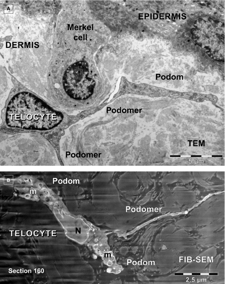
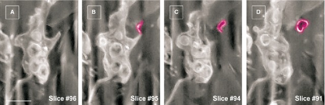
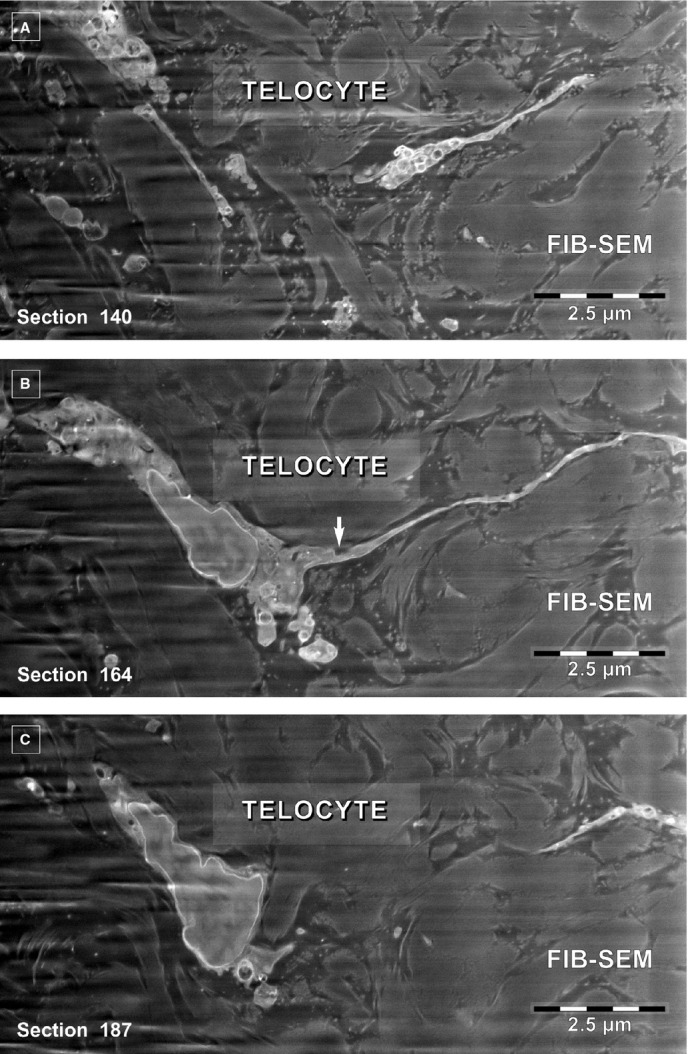
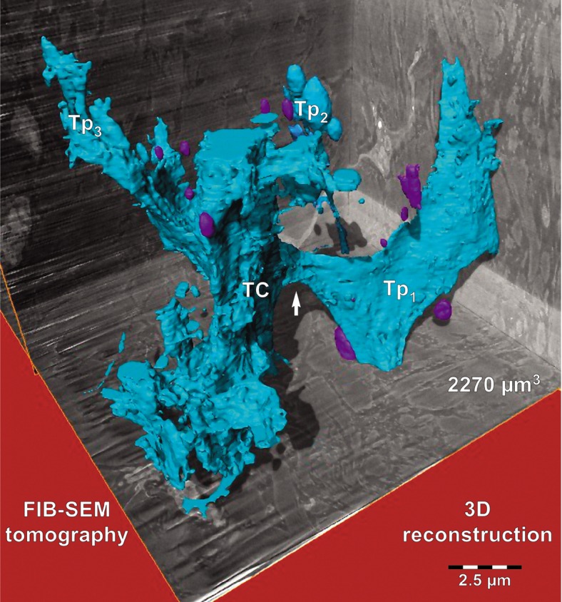
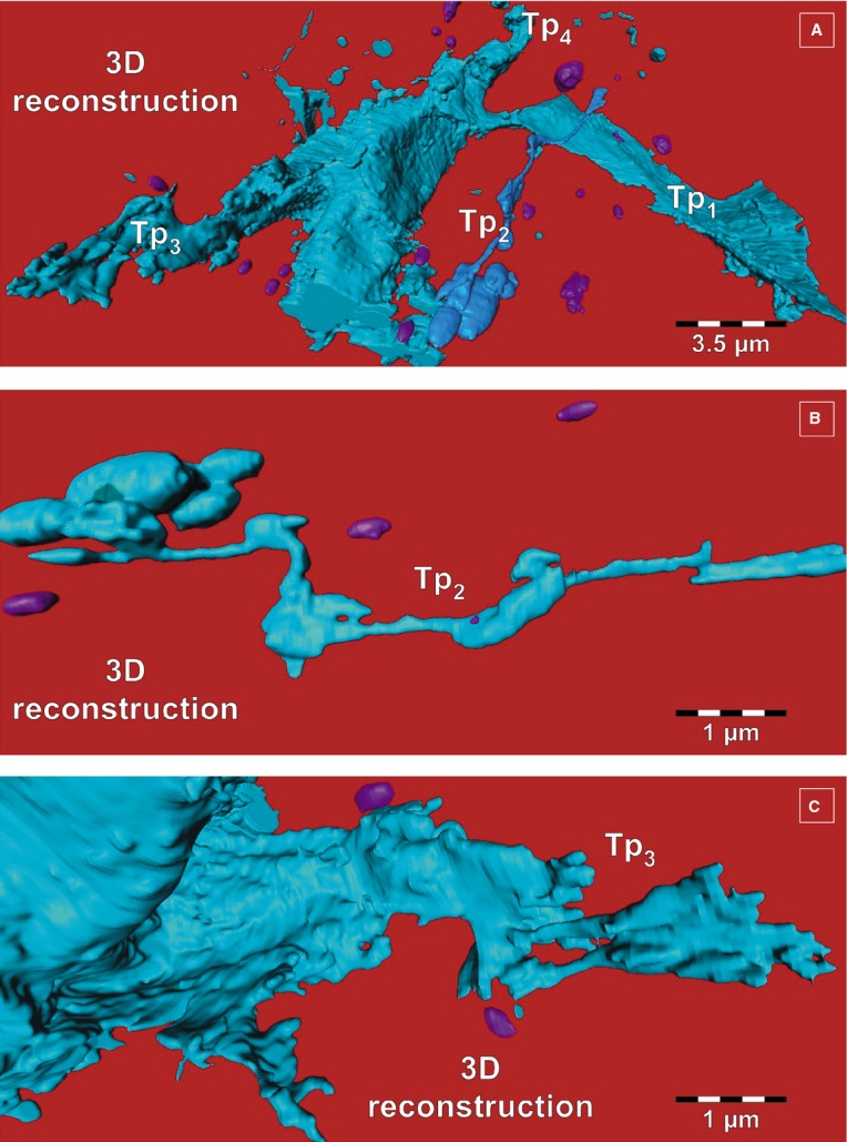
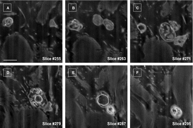
We have shown in 2012 the existence of telocytes (TCs) in human dermis. TCs were described by transmission electron microscopy (TEM) as interstitial cells located in non-epithelial spaces (stroma) of many organs (see www.telocytes.com). TCs have very long prolongations (tens to hundreds micrometers) named Telopodes (Tps). These Tps have a special conformation with dilated portions named podoms (containing mitochondria, endoplasmic reticulum and caveolae) and very thin segments (below resolving power of light microscopy), called podomers. To show the real 3D architecture of TC network, we used the most advanced available electron microscope technology: focused ion beam scanning electron microscopy (FIB-SEM) tomography. Generally, 3D reconstruction of dermal TCs by FIB-SEM tomography revealed the existence of Tps with various conformations: (i) long, flattened irregular veils (ribbon-like segments) with knobs, corresponding to podoms, and (ii) tubular structures (podomers) with uneven calibre because of irregular dilations (knobs) - the podoms. FIB-SEM tomography also showed numerous extracellular vesicles (diameter 438.6 ± 149.1 nm, n = 30) released by a human dermal TC. Our data might be useful for understanding the role(s) of TCs in intercellular signalling and communication, as well as for comprehension of pathologies like scleroderma, multiple sclerosis, psoriasis, etc.

摘要

2012年我们已证实人真皮中存在端粒细胞(TCs)。通过透射电子显微镜(TEM),端粒细胞被描述为位于许多器官非上皮空间(基质)的间质细胞(见www.telocytes.com)。端粒细胞具有名为端粒足(Tps)的非常长的延伸部分(数十至数百微米)。这些端粒足具有特殊的构象,有名为podom的扩张部分(含有线粒体、内质网和小窝)以及非常细的部分(低于光学显微镜的分辨能力),称为podomer。为了展示端粒细胞网络的真实三维结构,我们使用了现有的最先进电子显微镜技术:聚焦离子束扫描电子显微镜(FIB-SEM)断层扫描。一般来说,通过FIB-SEM断层扫描对真皮端粒细胞进行三维重建揭示了具有各种构象的端粒足的存在:(i)带有瘤状结构(对应于podom)的长而扁平的不规则面纱状(带状段),以及(ii)由于不规则扩张(瘤状结构)——即podom——而管径不均匀的管状结构(podomer)。FIB-SEM断层扫描还显示了人真皮端粒细胞释放的大量细胞外囊泡(直径438.6±149.1纳米,n = 30)。我们的数据可能有助于理解端粒细胞在细胞间信号传导和通讯中的作用,以及有助于理解诸如硬皮病、多发性硬化症、牛皮癣等疾病。

https://cdn.ncbi.nlm.nih.gov/pmc/blobs/f347/4395186/1c6796f15541/jcmm0019-0714-f6.jpg
https://cdn.ncbi.nlm.nih.gov/pmc/blobs/f347/4395186/3289820a1966/jcmm0019-0714-f1.jpg
https://cdn.ncbi.nlm.nih.gov/pmc/blobs/f347/4395186/b82f3ffd0bfc/jcmm0019-0714-f2.jpg
https://cdn.ncbi.nlm.nih.gov/pmc/blobs/f347/4395186/cf3504d278b8/jcmm0019-0714-f3.jpg
https://cdn.ncbi.nlm.nih.gov/pmc/blobs/f347/4395186/236aaf5d2a6c/jcmm0019-0714-f4.jpg
https://cdn.ncbi.nlm.nih.gov/pmc/blobs/f347/4395186/ce2010304754/jcmm0019-0714-f5.jpg
https://cdn.ncbi.nlm.nih.gov/pmc/blobs/f347/4395186/1c6796f15541/jcmm0019-0714-f6.jpg

相似文献

1
FIB-SEM tomography of human skin telocytes and their extracellular vesicles.人皮肤端细胞及其细胞外囊泡的聚焦离子束扫描电子显微镜断层扫描
J Cell Mol Med. 2015 Apr;19(4):714-22. doi: 10.1111/jcmm.12578.
2
Ligamentum arteriosum and its telocytes: An ultrastructure description.动脉韧带及其间充质干细胞:超微结构描述。
Anat Rec (Hoboken). 2023 Jan;306(1):187-192. doi: 10.1002/ar.25052. Epub 2022 Aug 6.
3
The Cutaneous Telocytes.皮肤间充质干细胞。 (备注:原文中“Telocytes”常见释义为“间充质干细胞” ,但结合专业语境,这里可能翻译为“皮肤特络细胞”更准确,你可根据实际情况判断。此次按照指令要求先给出这个译文供你参考。) 正确译文:皮肤特络细胞
Adv Exp Med Biol. 2016;913:303-323. doi: 10.1007/978-981-10-1061-3_20.
4
Human cardiac telocytes: 3D imaging by FIB-SEM tomography.人类心脏间质细胞:通过聚焦离子束扫描电子显微镜断层扫描进行三维成像
J Cell Mol Med. 2014 Nov;18(11):2157-64. doi: 10.1111/jcmm.12468. Epub 2014 Oct 17.
5
Telocytes: novel interstitial cells present in the testis parenchyma of the Chinese soft-shelled turtle Pelodiscus sinensis.端细胞:中华鳖睾丸实质中存在的新型间质细胞。
J Cell Mol Med. 2015 Dec;19(12):2888-99. doi: 10.1111/jcmm.12731.
6
The Third Dimension of Telocytes Revealed by FIB-SEM Tomography.聚焦离子束扫描电子显微镜断层扫描揭示的间质细胞的三维结构
Adv Exp Med Biol. 2016;913:325-334. doi: 10.1007/978-981-10-1061-3_21.
7
Effect of Melatonin on Telocytes in the Seminal Vesicle of the Soay Ram: An Immunohistochemical, Ultrastructural and Morphometrical Study.褪黑素对索艾羊精囊间质细胞的影响:一项免疫组织化学、超微结构及形态计量学研究
Cells Tissues Organs. 2017;203(1):29-54. doi: 10.1159/000449500. Epub 2016 Nov 2.
8
Transmission electron microscope evidence of telocytes in canine dura mater.犬硬脑膜中存在端细胞的透射电子显微镜证据。
J Cell Mol Med. 2016 Jan;20(1):188-92. doi: 10.1111/jcmm.12726.
9
CD34 stromal cells/telocytes and their role in mouse lung development: Light microscopy, immunofluorescence, ultrastructural and scanning electron microscopy evidence.CD34 基质细胞/间质细胞及其在小鼠肺发育中的作用:光镜、免疫荧光、超微结构和扫描电镜证据。
Cell Biol Int. 2024 Nov;48(11):1680-1697. doi: 10.1002/cbin.12223. Epub 2024 Aug 4.
10
Telopodes of telocytes are influenced in vitro by redox conditions and ageing.端粒细胞的端粒在体外受氧化还原条件和衰老的影响。
Mol Cell Biochem. 2015 Dec;410(1-2):165-74. doi: 10.1007/s11010-015-2548-2. Epub 2015 Sep 3.

引用本文的文献

1
DeepSCEM: A User-Friendly Solution for Deep Learning-Based Image Segmentation in Cellular Electron Microscopy.DeepSCEM:细胞电子显微镜中基于深度学习的图像分割的用户友好解决方案。
Biol Cell. 2025 Sep;117(9):e70032. doi: 10.1111/boc.70032.
2
Telocytes: history, origin, identification, structure, distribution, and functions.间充质干细胞:历史、起源、鉴定、结构、分布及功能。
Histochem Cell Biol. 2025 Aug 29;163(1):86. doi: 10.1007/s00418-025-02413-1.
3
Skin Telocyte Secretome as Conditioned Medium Prevents Profibrotic Differentiation of Skin Fibroblasts into Myofibroblasts.

本文引用的文献

1
Uterine Telocytes: A Review of Current Knowledge.子宫间充质干细胞:当前知识综述。
Biol Reprod. 2015 Jul;93(1):10. doi: 10.1095/biolreprod.114.125906. Epub 2015 Feb 18.
2
Cardiac telocytes and fibroblasts in primary culture: different morphologies and immunophenotypes.原代培养的心脏间充质干细胞和成纤维细胞:不同的形态和免疫表型。
PLoS One. 2015 Feb 18;10(2):e0115991. doi: 10.1371/journal.pone.0115991. eCollection 2015.
3
Towards the imaging of Weibel-Palade body biogenesis by serial block face-scanning electron microscopy.
作为条件培养基的皮肤间充质干细胞分泌组可防止皮肤成纤维细胞向肌成纤维细胞的促纤维化分化。
Int J Mol Sci. 2025 Feb 2;26(3):1284. doi: 10.3390/ijms26031284.
4
Descriptive embryological insights of the colorectum of quail embryos with concern to its functional morphology.探讨禽类胚胎直肠的胚胎发生学特点及其与功能形态学的关系。
BMC Vet Res. 2024 Nov 6;20(1):508. doi: 10.1186/s12917-024-04341-z.
5
Developmental characteristics of cutaneous telocytes in late embryos of the silky fowl.丝羽乌骨鸡晚期胚胎皮肤中的结缔组织细胞的发育特征。
Eur J Histochem. 2024 Oct 15;68(4):4089. doi: 10.4081/ejh.2024.4089.
6
Telocytes: current methods of research, challenges and future perspectives.间质细胞:当前的研究方法、挑战与未来展望。
Cell Tissue Res. 2024 May;396(2):141-155. doi: 10.1007/s00441-024-03888-5. Epub 2024 Mar 28.
7
Telocytes/CD34+ Stromal Cells in the Normal, Hyperplastic, and Adenomatous Human Parathyroid Glands.正常、增生和腺瘤性人甲状旁腺中的 Telocytes/CD34+基质细胞。
Int J Mol Sci. 2023 Jul 28;24(15):12118. doi: 10.3390/ijms241512118.
8
Identification and protective role of CD34 stromal cells/telocytes in experimental autoimmune encephalomyelitis (EAE) mouse spleen.鉴定和保护作用的 CD34 基质细胞/间质细胞在实验性自身免疫性脑脊髓炎 (EAE) 鼠脾。
Histochem Cell Biol. 2023 Jul;160(1):11-25. doi: 10.1007/s00418-023-02186-5. Epub 2023 Apr 4.
9
Delimiting CD34+ Stromal Cells/Telocytes Are Resident Mesenchymal Cells That Participate in Neovessel Formation in Skin Kaposi Sarcoma.CD34+ 基质细胞/间质细胞是定位于皮肤卡波西肉瘤中的固有间充质细胞,参与新血管形成。
Int J Mol Sci. 2023 Feb 14;24(4):3793. doi: 10.3390/ijms24043793.
10
Skin cell-derived extracellular vesicles: a promising therapeutic strategy for cutaneous injury.皮肤细胞衍生的细胞外囊泡:一种有前景的皮肤损伤治疗策略。
Burns Trauma. 2022 Oct 18;10:tkac037. doi: 10.1093/burnst/tkac037. eCollection 2022.
通过连续块面扫描电子显微镜对魏贝尔-帕拉德小体生物发生进行成像研究。
J Microsc. 2015 Aug;259(2):97-104. doi: 10.1111/jmi.12222. Epub 2015 Jan 23.
4
Quantitative characterization of endothelial cell morphologies depending on shear stress in different blood vessels of domestic pigs using a focused ion beam and high resolution scanning electron microscopy (FIB-SEM).使用聚焦离子束和高分辨率扫描电子显微镜(FIB-SEM)对家猪不同血管中依赖于剪切应力的内皮细胞形态进行定量表征。
Tissue Cell. 2015 Apr;47(2):205-12. doi: 10.1016/j.tice.2014.12.005. Epub 2014 Dec 26.
5
Telocytes transfer extracellular vesicles loaded with microRNAs to stem cells.间充质干细胞将装载有微小核糖核酸的细胞外囊泡转移至干细胞。
J Cell Mol Med. 2015 Feb;19(2):351-8. doi: 10.1111/jcmm.12529. Epub 2015 Jan 20.
6
New developments in electron microscopy for serial image acquisition of neuronal profiles.用于神经元轮廓连续图像采集的电子显微镜新进展。
Microscopy (Oxf). 2015 Feb;64(1):27-36. doi: 10.1093/jmicro/dfu111. Epub 2015 Jan 5.
7
Telocytes and putative stem cells in ageing human heart.衰老人类心脏中的间质细胞和假定的干细胞。
J Cell Mol Med. 2015 Jan;19(1):31-45. doi: 10.1111/jcmm.12509. Epub 2014 Dec 25.
8
Minimal experimental requirements for definition of extracellular vesicles and their functions: a position statement from the International Society for Extracellular Vesicles.最小实验要求定义细胞外囊泡及其功能:国际细胞外囊泡学会的立场声明。
J Extracell Vesicles. 2014 Dec 22;3:26913. doi: 10.3402/jev.v3.26913. eCollection 2014.
9
Extracellular vesicles: potential roles in regenerative medicine.细胞外囊泡:在再生医学中的潜在作用。
Front Immunol. 2014 Dec 3;5:608. doi: 10.3389/fimmu.2014.00608. eCollection 2014.
10
Investigation of resins suitable for the preparation of biological sample for 3-D electron microscopy.适用于制备用于三维电子显微镜检查的生物样品的树脂研究。
J Struct Biol. 2015 Feb;189(2):135-46. doi: 10.1016/j.jsb.2014.10.009. Epub 2014 Nov 26.