• 文献检索
  • 文档翻译
  • 深度研究
  • 学术资讯
  • Suppr Zotero 插件Zotero 插件
  • 邀请有礼
  • 套餐&价格
  • 历史记录
应用&插件
Suppr Zotero 插件Zotero 插件浏览器插件Mac 客户端Windows 客户端微信小程序
定价
高级版会员购买积分包购买API积分包
服务
文献检索文档翻译深度研究API 文档MCP 服务
关于我们
关于 Suppr公司介绍联系我们用户协议隐私条款
关注我们

Suppr 超能文献

核心技术专利:CN118964589B侵权必究
粤ICP备2023148730 号-1Suppr @ 2026

文献检索

告别复杂PubMed语法,用中文像聊天一样搜索,搜遍4000万医学文献。AI智能推荐,让科研检索更轻松。

立即免费搜索

文件翻译

保留排版,准确专业,支持PDF/Word/PPT等文件格式,支持 12+语言互译。

免费翻译文档

深度研究

AI帮你快速写综述,25分钟生成高质量综述,智能提取关键信息,辅助科研写作。

立即免费体验

通过连续块面扫描电子显微镜对魏贝尔-帕拉德小体生物发生进行成像研究。

Towards the imaging of Weibel-Palade body biogenesis by serial block face-scanning electron microscopy.

作者信息

Mourik M J, Faas F G A, Zimmermann H, Eikenboom J, Koster A J

机构信息

Department of Molecular Cell Biology, Leiden University Medical Center, Leiden, the Netherlands.

Carl Zeiss Microscopy GmbH, Munich, Germany.

出版信息

J Microsc. 2015 Aug;259(2):97-104. doi: 10.1111/jmi.12222. Epub 2015 Jan 23.

DOI:10.1111/jmi.12222
PMID:25644989
原文链接:https://pmc.ncbi.nlm.nih.gov/articles/PMC4670698/
Abstract

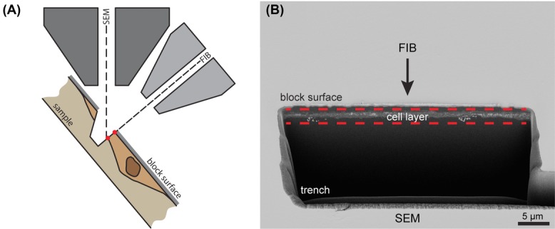
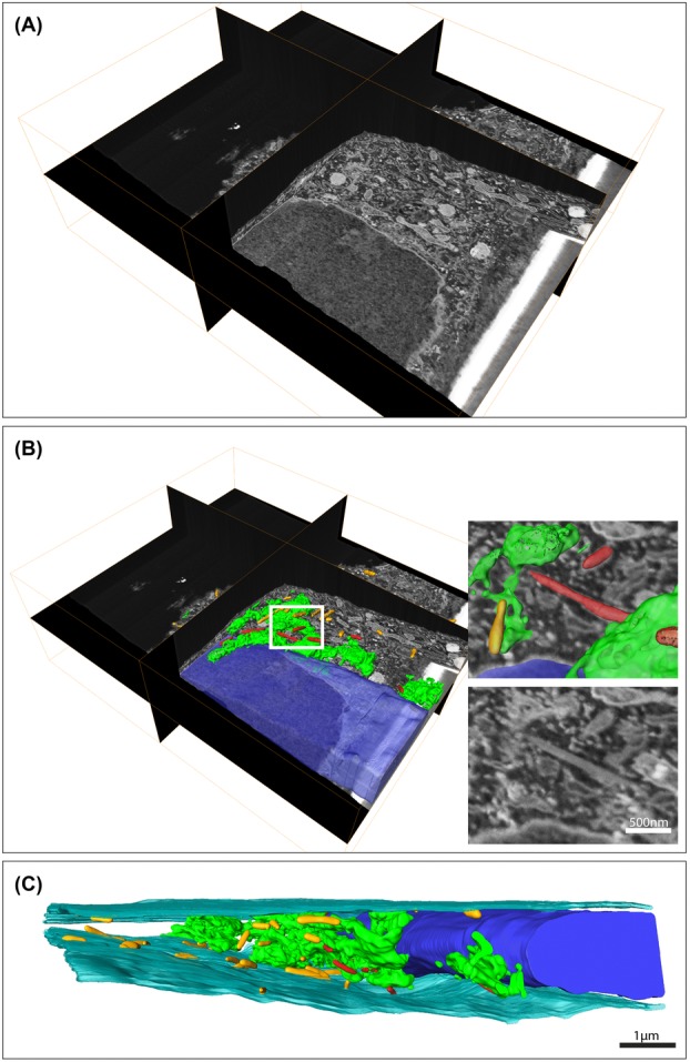
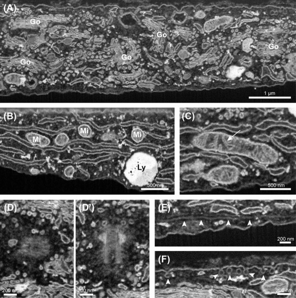
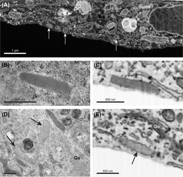
Electron microscopy is used in biological research to study the ultrastructure at high resolution to obtain information on specific cellular processes. Serial block face-scanning electron microscopy is a relatively novel electron microscopy imaging technique that allows three-dimensional characterization of the ultrastructure in both tissues and cells by measuring volumes of thousands of cubic micrometres yet at nanometre-scale resolution. In the scanning electron microscope, repeatedly an image is acquired followed by the removal of a thin layer resin embedded biological material by either a microtome or a focused ion beam. In this way, each recorded image contains novel structural information which can be used for three-dimensional analysis. Here, we explore focused ion beam facilitated serial block face-scanning electron microscopy to study the endothelial cell-specific storage organelles, the Weibel-Palade bodies, during their biogenesis at the Golgi apparatus. Weibel-Palade bodies predominantly contain the coagulation protein Von Willebrand factor which is secreted by the cell upon vascular damage. Using focused ion beam facilitated serial block face-scanning electron microscopy we show that the technique has the sensitivity to clearly reveal subcellular details like mitochondrial cristae and small vesicles with a diameter of about 50 nm. Also, we reveal numerous associations between Weibel-Palade bodies and Golgi stacks which became conceivable in large-scale three-dimensional data. We demonstrate that serial block face-scanning electron microscopy is a promising tool that offers an alternative for electron tomography to study subcellular organelle interactions in the context of a complete cell.

摘要

电子显微镜在生物学研究中用于以高分辨率研究超微结构,以获取有关特定细胞过程的信息。连续块面扫描电子显微镜是一种相对新颖的电子显微镜成像技术,它能够通过测量数千立方微米的体积,但以纳米级分辨率对组织和细胞中的超微结构进行三维表征。在扫描电子显微镜中,反复采集图像,然后用切片机或聚焦离子束去除嵌入生物材料的薄层树脂。通过这种方式,每个记录的图像都包含可用于三维分析的新结构信息。在这里,我们探索聚焦离子束辅助的连续块面扫描电子显微镜,以研究内皮细胞特异性储存细胞器——魏-帕小体在高尔基体生物发生过程中的情况。魏-帕小体主要含有凝血蛋白血管性血友病因子,该因子在血管损伤时由细胞分泌。使用聚焦离子束辅助的连续块面扫描电子显微镜,我们表明该技术具有清晰揭示亚细胞细节的敏感性,如线粒体嵴和直径约50nm的小泡。此外,我们揭示了魏-帕小体与高尔基体堆栈之间的大量关联,这在大规模三维数据中是可以想象的。我们证明,连续块面扫描电子显微镜是一种很有前途的工具,为电子断层扫描提供了一种替代方法,用于在完整细胞的背景下研究亚细胞器相互作用。

https://cdn.ncbi.nlm.nih.gov/pmc/blobs/b331/4670698/e34b3e8304e2/jmi0259-0097-f4.jpg
https://cdn.ncbi.nlm.nih.gov/pmc/blobs/b331/4670698/6864c6c34762/jmi0259-0097-f1.jpg
https://cdn.ncbi.nlm.nih.gov/pmc/blobs/b331/4670698/11b1597888d0/jmi0259-0097-f2.jpg
https://cdn.ncbi.nlm.nih.gov/pmc/blobs/b331/4670698/ee58c79a719f/jmi0259-0097-f3.jpg
https://cdn.ncbi.nlm.nih.gov/pmc/blobs/b331/4670698/e34b3e8304e2/jmi0259-0097-f4.jpg

相似文献

1
Towards the imaging of Weibel-Palade body biogenesis by serial block face-scanning electron microscopy.通过连续块面扫描电子显微镜对魏贝尔-帕拉德小体生物发生进行成像研究。
J Microsc. 2015 Aug;259(2):97-104. doi: 10.1111/jmi.12222. Epub 2015 Jan 23.
2
Three-dimensional shape of the Golgi apparatus in different cell types: serial section scanning electron microscopy of the osmium-impregnated Golgi apparatus.不同细胞类型中高尔基体的三维形态:锇浸渍高尔基体的连续切片扫描电子显微镜观察
Microscopy (Oxf). 2016 Apr;65(2):145-57. doi: 10.1093/jmicro/dfv360. Epub 2015 Nov 24.
3
Developing 3D SEM in a broad biological context.在广泛的生物学背景下开发三维扫描电子显微镜。
J Microsc. 2015 Aug;259(2):80-96. doi: 10.1111/jmi.12211. Epub 2015 Jan 26.
4
Content delivery to newly forming Weibel-Palade bodies is facilitated by multiple connections with the Golgi apparatus.内容递送到新形成的 Weibel-Palade 小体是通过与高尔基体的多个连接来实现的。
Blood. 2015 May 28;125(22):3509-16. doi: 10.1182/blood-2014-10-608596. Epub 2015 Feb 25.
5
New developments in electron microscopy for serial image acquisition of neuronal profiles.用于神经元轮廓连续图像采集的电子显微镜新进展。
Microscopy (Oxf). 2015 Feb;64(1):27-36. doi: 10.1093/jmicro/dfu111. Epub 2015 Jan 5.
6
FIB-SEM tomography in biology.生物学中的聚焦离子束扫描电子显微镜断层成像
Methods Mol Biol. 2014;1117:541-58. doi: 10.1007/978-1-62703-776-1_24.
7
Volume scanning electron microscopy for imaging biological ultrastructure.用于生物超微结构成像的体积扫描电子显微镜。
Biol Cell. 2016 Nov;108(11):307-323. doi: 10.1111/boc.201600024. Epub 2016 Aug 12.
8
Three-dimensional imaging of adherent cells using FIB/SEM and STEM.使用聚焦离子束/扫描电子显微镜(FIB/SEM)和扫描透射电子显微镜(STEM)对贴壁细胞进行三维成像。
Methods Mol Biol. 2014;1117:617-38. doi: 10.1007/978-1-62703-776-1_27.
9
Weibel-Palade bodies recruit Rab27 by a content-driven, maturation-dependent mechanism that is independent of cell type.魏贝尔-帕拉德小体通过一种不依赖细胞类型、由内容物驱动且依赖成熟的机制募集Rab27。
J Cell Sci. 2003 Oct 1;116(Pt 19):3939-48. doi: 10.1242/jcs.00711. Epub 2003 Aug 19.
10
Correlative light microscopy and electron tomography to study Von Willebrand factor exocytosis from vascular endothelial cells.利用相关光学显微镜和电子断层扫描技术研究血管内皮细胞中血管性血友病因子的胞吐作用。
Methods Cell Biol. 2014;124:71-92. doi: 10.1016/B978-0-12-801075-4.00004-5.

引用本文的文献

1
Comparison of 3D cellular imaging techniques based on scanned electron probes: Serial block face SEM vs. Axial bright-field STEM tomography.基于扫描电子探针的 3D 细胞成像技术比较:连续块面扫描电镜与轴向明场扫描透射电镜断层成像。
J Struct Biol. 2018 Jun;202(3):216-228. doi: 10.1016/j.jsb.2018.01.012. Epub 2018 Feb 1.
2
Conductive resins improve charging and resolution of acquired images in electron microscopic volume imaging.导电树脂可提高电子显微镜体积成像中获取图像的充电和分辨率。
Sci Rep. 2016 Mar 29;6:23721. doi: 10.1038/srep23721.
3
FIB-SEM tomography of human skin telocytes and their extracellular vesicles.

本文引用的文献

1
A two-tier Golgi-based control of organelle size underpins the functional plasticity of endothelial cells.双层高尔基基体控制细胞器大小,为内皮细胞的功能可塑性提供基础。
Dev Cell. 2014 May 12;29(3):292-304. doi: 10.1016/j.devcel.2014.03.021. Epub 2014 May 1.
2
Correlative 3D imaging: CLSM and FIB-SEM tomography using high-pressure frozen, freeze-substituted biological samples.相关三维成像:使用高压冷冻、冷冻置换生物样品的共聚焦激光扫描显微镜(CLSM)和聚焦离子束扫描电子显微镜(FIB-SEM)断层扫描
Methods Mol Biol. 2014;1117:593-616. doi: 10.1007/978-1-62703-776-1_26.
3
Is EM dead?
人皮肤端细胞及其细胞外囊泡的聚焦离子束扫描电子显微镜断层扫描
J Cell Mol Med. 2015 Apr;19(4):714-22. doi: 10.1111/jcmm.12578.
急诊医学是否已死?
J Cell Sci. 2013 Oct 15;126(Pt 20):4545-52. doi: 10.1242/jcs.124123.
4
Synaptic inputs compete during rapid formation of the calyx of Held: a new model system for neural development.在快速形成 Held 触须的过程中,突触输入相互竞争:一个用于神经发育的新模型系统。
J Neurosci. 2013 Aug 7;33(32):12954-69. doi: 10.1523/JNEUROSCI.1087-13.2013.
5
FIB/SEM tomography with TEM-like resolution for 3D imaging of high-pressure frozen cells.采用类似于 TEM 的分辨率的 FIB/SEM 断层扫描技术对高压冷冻细胞进行 3D 成像。
Histochem Cell Biol. 2012 Oct;138(4):549-56. doi: 10.1007/s00418-012-1020-6. Epub 2012 Aug 25.
6
Correlative light-electron microscopy a potent tool for the imaging of rare or unique cellular and tissue events and structures.相关光电子显微镜是一种用于对罕见或独特的细胞及组织事件与结构进行成像的强大工具。
Methods Enzymol. 2012;504:201-19. doi: 10.1016/B978-0-12-391857-4.00010-0.
7
High-contrast en bloc staining of neuronal tissue for field emission scanning electron microscopy.高对比度整块染色的神经元组织用于场发射扫描电子显微镜。
Nat Protoc. 2012 Jan 12;7(2):193-206. doi: 10.1038/nprot.2011.439.
8
Modern electron microscopy methods for C. elegans.用于秀丽隐杆线虫的现代电子显微镜方法。
Methods Cell Biol. 2012;107:93-149. doi: 10.1016/B978-0-12-394620-1.00004-7.
9
Correlative 3D imaging of whole mammalian cells with light and electron microscopy.用光镜和电镜对全哺乳动物细胞进行相关的三维成像。
J Struct Biol. 2011 Dec;176(3):268-78. doi: 10.1016/j.jsb.2011.08.013. Epub 2011 Sep 1.
10
Functional architecture of Weibel-Palade bodies.Weibel-Palade bodies 的功能结构。
Blood. 2011 May 12;117(19):5033-43. doi: 10.1182/blood-2010-09-267492. Epub 2011 Jan 25.