Seyed Foroutan Kamal, Khodarahmi Ali, Alavi Hootan, Pedram Sepehr, Baghaban Eslaminejad Mohamad Reza, Bordbar Sima
Department of Plastic Surgery, Hazrat Fatemeh Hospital, Iran University of Medical Sciences, Tehran, IR Iran.
Department of Plastic Surgery, Bahonar Hospital, Kerman University of Medical Sciences, Kerman, IR Iran.
Trauma Mon. 2015 Feb;20(1):e23325. doi: 10.5812/traumamon.23325. Epub 2015 Jan 17.
Peripheral nerve repair with sufficient functional recovery is an important issue in reconstructive surgery. Stem cells have attracted extensive research interest in recent years.
The purpose of this study was to compare the vein conduit technique, with and without the addition of mesenchymal stem cells in gap-less nerve injury repair in rats.
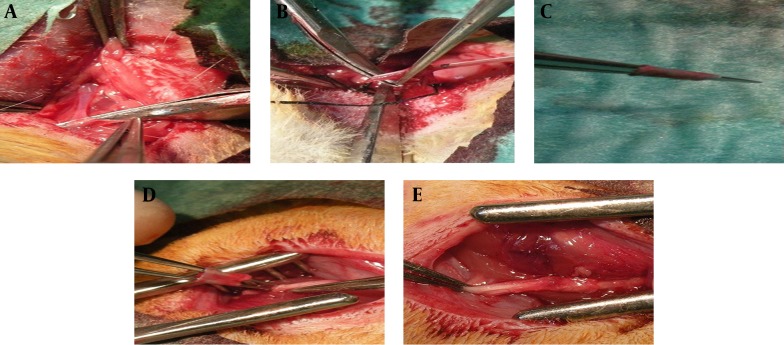
In this study, 36 Wistar rats were randomly allocated to three groups: In the first group, nerve repair was performed with simple neurorrhaphy (control group), in the second group, nerve repair was done with vein conduit over site (vein conduit group) and in the third group, bone marrow stem cells were instilled into the vein conduit (stem cell group) after nerve repair with vein conduit over site. Six weeks after the intervention, the sciatic function index, electrophysiological study and histological examination were performed.
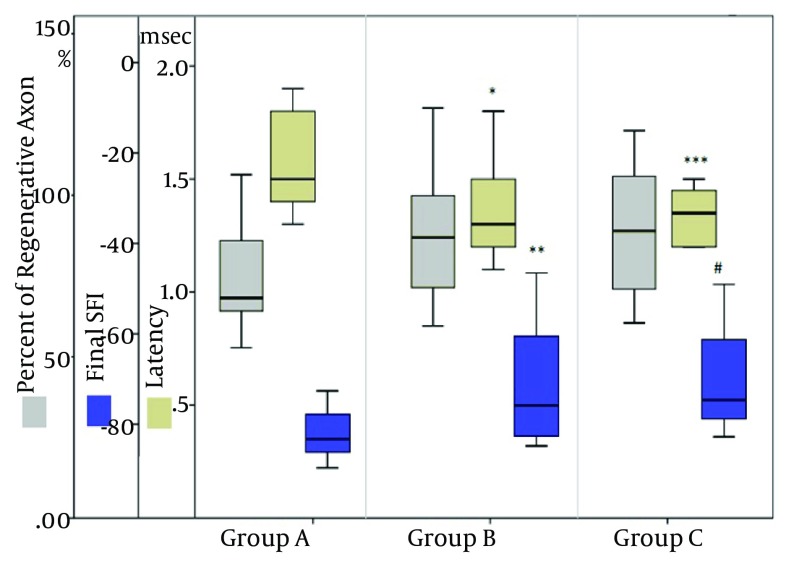
All animals tolerated the surgical procedures and survived well. The sciatic function index and latency were significantly improved in the vein conduit (P = 0.04 and 0.03, respectively) and stem cell group (P = 0.02 and 0.03, respectively) compared with the control group. No significant difference was observed in sciatic function and latency between the vein conduit and stem-cell groups. Moreover, histological analysis showed no significant difference in regenerative density between these two groups.
The results of this study showed that the meticulous microsurgical nerve repair, which was performed using the vein tubulization induced significantly better sciatic nerve regeneration. However, the addition of bone marrow mesenchymal stem cell to vein conduit failed to promote any significant changes in regeneration outcome.
在重建手术中,实现具有足够功能恢复的周围神经修复是一个重要问题。近年来,干细胞吸引了广泛的研究兴趣。
本研究旨在比较在大鼠无间隙神经损伤修复中,添加和不添加间充质干细胞的静脉导管技术。
在本研究中,36只Wistar大鼠被随机分为三组:第一组采用单纯神经缝合进行神经修复(对照组),第二组采用静脉导管覆盖部位进行神经修复(静脉导管组),第三组在采用静脉导管覆盖部位进行神经修复后,将骨髓干细胞注入静脉导管(干细胞组)。干预6周后,进行坐骨神经功能指数、电生理研究和组织学检查。
所有动物均耐受手术操作且存活良好。与对照组相比,静脉导管组(分别为P = 0.04和0.03)和干细胞组(分别为P = 0.02和0.03)的坐骨神经功能指数和潜伏期均有显著改善。静脉导管组和干细胞组之间在坐骨神经功能和潜伏期方面未观察到显著差异。此外,组织学分析显示这两组之间在再生密度方面无显著差异。
本研究结果表明,使用静脉管道化进行的精细显微外科神经修复可显著促进坐骨神经再生。然而,在静脉导管中添加骨髓间充质干细胞未能促进再生结果发生任何显著变化。