• 文献检索
  • 文档翻译
  • 深度研究
  • 学术资讯
  • Suppr Zotero 插件Zotero 插件
  • 邀请有礼
  • 套餐&价格
  • 历史记录
应用&插件
Suppr Zotero 插件Zotero 插件浏览器插件Mac 客户端Windows 客户端微信小程序
定价
高级版会员购买积分包购买API积分包
服务
文献检索文档翻译深度研究API 文档MCP 服务
关于我们
关于 Suppr公司介绍联系我们用户协议隐私条款
关注我们

Suppr 超能文献

核心技术专利:CN118964589B侵权必究
粤ICP备2023148730 号-1Suppr @ 2026

文献检索

告别复杂PubMed语法,用中文像聊天一样搜索,搜遍4000万医学文献。AI智能推荐,让科研检索更轻松。

立即免费搜索

文件翻译

保留排版,准确专业,支持PDF/Word/PPT等文件格式,支持 12+语言互译。

免费翻译文档

深度研究

AI帮你快速写综述,25分钟生成高质量综述,智能提取关键信息,辅助科研写作。

立即免费体验

贝伐单抗治疗复杂性后极部黑素细胞瘤。

Bevacizumab for the treatment of a complicated posterior melanocytoma.

作者信息

Urrets-Zavalia Julio A, Crim Nicolas, Esposito Evangelina, Correa Leandro, Gonzalez-Castellanos M Eugenia, Martinez Dana

机构信息

Department of Ophthalmology, University Clinic Reina Fabiola, Universidad Catolica de Cordoba, Cordoba, Argentina.

出版信息

Clin Ophthalmol. 2015 Mar 6;9:455-9. doi: 10.2147/OPTH.S80152. eCollection 2015.

DOI:10.2147/OPTH.S80152
PMID:25834382
原文链接:https://pmc.ncbi.nlm.nih.gov/articles/PMC4358416/
Abstract

PURPOSE

To present a case of a complicated posterior melanocytoma that was successfully treated with intravitreal bevacizumab.

CASE REPORT

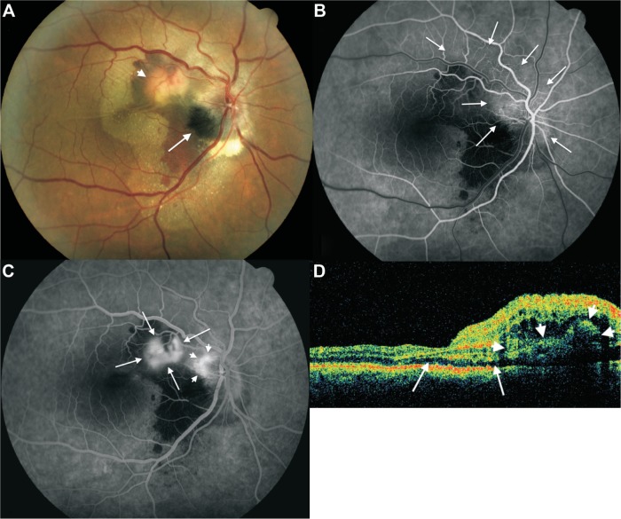
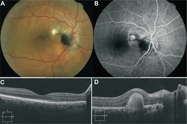
A 50-year-old Caucasian man was referred with sudden-onset metamorphopsia and decreased vision in his right eye over the course of the last 2 months. His best-corrected visual acuity was 20/80 and poorer than Jaeger 14 in the right eye, and 20/20 and Jaeger 1 in his left eye. In the right fundus, there was a melanocytic lesion occupying the inferotemporal quadrant of the optic disk, extending to the adjacent choroid inferiorly; optic nerve edema, superotemporal retinal vein dilatation, and subretinal fluid under the macula and nasal half of the posterior pole were observed, and a subretinal choroidal neovascularization complex was observed adjacent to the superotemporal margin of the optic disk, confirmed by fluorescein angiography, surrounded by a dense subretinal hemorrhage. Optical coherence tomography showed retinal edema and detachment of neurosensory retina. The patient was treated with three consecutive doses on a monthly basis of intravitreal 1.25 mg/0.05 mL bevacizumab. Visual acuity recovered rapidly, and at 4 months after treatment, it was 20/20 and Jaeger 1, with complete resolution of macular edema and subretinal fluid and hemorrhage. After 3 years of follow-up, best-corrected visual acuity remained stable, macular area was normal, and there was no evident optic nerve edema, retinal vein caliber and aspect were normal, and there was no significant change of the tumor. Fluorescein angiography only evidenced late staining of choroidal neovascularization scar, and optical coherence tomography showed a normal macular anatomy.

CONCLUSION

Intravitreal bevacizumab was effective in the treatment of choroidal neovascularization, optic nerve edema, venous dilatation, and local capillary telangiectasia, complicating an optic disk melanocytoma.

摘要

目的

报告1例采用玻璃体内注射贝伐单抗成功治疗的复杂性视盘黑色素细胞瘤病例。

病例报告

一名50岁的白种男性在过去2个月内右眼突然出现视物变形和视力下降,遂前来就诊。其右眼最佳矫正视力为20/80,低于耶格视力表14级;左眼最佳矫正视力为20/20,耶格视力表1级。右眼眼底可见一个黑色素细胞病变,占据视盘颞下象限,并向下延伸至相邻脉络膜;可见视神经水肿、颞上视网膜静脉扩张、黄斑及后极部鼻侧一半的视网膜下液,且在视盘颞上缘附近可见一个视网膜下脉络膜新生血管复合体,荧光素血管造影证实该复合体周围有密集的视网膜下出血。光学相干断层扫描显示视网膜水肿及神经感觉层视网膜脱离。该患者每月连续3次接受玻璃体内注射1.25 mg/0.05 mL贝伐单抗治疗。视力迅速恢复,治疗4个月后,视力为20/20,耶格视力表1级,黄斑水肿、视网膜下液及出血完全消退。随访3年后,最佳矫正视力保持稳定,黄斑区正常,无明显视神经水肿,视网膜静脉管径及外观正常,肿瘤无明显变化。荧光素血管造影仅显示脉络膜新生血管瘢痕的晚期染色,光学相干断层扫描显示黄斑解剖结构正常。

结论

玻璃体内注射贝伐单抗可有效治疗视盘黑色素细胞瘤合并的脉络膜新生血管、视神经水肿、静脉扩张及局部毛细血管扩张。

https://cdn.ncbi.nlm.nih.gov/pmc/blobs/63d5/4358416/dbbf24766c34/opth-9-455Fig2.jpg
https://cdn.ncbi.nlm.nih.gov/pmc/blobs/63d5/4358416/b67130fe35b1/opth-9-455Fig1.jpg
https://cdn.ncbi.nlm.nih.gov/pmc/blobs/63d5/4358416/dbbf24766c34/opth-9-455Fig2.jpg
https://cdn.ncbi.nlm.nih.gov/pmc/blobs/63d5/4358416/b67130fe35b1/opth-9-455Fig1.jpg
https://cdn.ncbi.nlm.nih.gov/pmc/blobs/63d5/4358416/dbbf24766c34/opth-9-455Fig2.jpg

相似文献

1
Bevacizumab for the treatment of a complicated posterior melanocytoma.贝伐单抗治疗复杂性后极部黑素细胞瘤。
Clin Ophthalmol. 2015 Mar 6;9:455-9. doi: 10.2147/OPTH.S80152. eCollection 2015.
2
Intravitreal Bevacizumab for Traumatic Choroidal Rupture.玻璃体内注射贝伐单抗治疗外伤性脉络膜破裂
Optom Vis Sci. 2015 Oct;92(10):e363-7. doi: 10.1097/OPX.0000000000000677.
3
Choroidal neovascularization of optic disk melanocytoma treated with bevacizumab.贝伐单抗治疗视盘黑素细胞瘤的脉络膜新生血管形成
Eur J Ophthalmol. 2012 May-Jun;22(3):503-5. doi: 10.5301/ejo.5000060.
4
Optic disk melanocytoma associated with polypoidal choroidal vasculopathy lesions, after combination treatment of photodynamic therapy and intavitreal aflibercept (Eylea), a case report.光动力疗法联合玻璃体内注射阿柏西普(阿瓦斯汀)治疗后,视盘黑素细胞瘤合并息肉状脉络膜血管病变:一例报告
BMC Ophthalmol. 2018 Oct 12;18(1):267. doi: 10.1186/s12886-018-0927-7.
5
Successful treatment of choroidal neovascularization complicating a choroidal osteoma with intravitreal bevacizumab.玻璃体内注射贝伐单抗成功治疗脉络膜骨瘤合并的脉络膜新生血管。
Retin Cases Brief Rep. 2010 Fall;4(4):303-5. doi: 10.1097/ICB.0b013e3181aff523.
6
Intravitreal ranibizumab and bevacizumab therapy for choroidal neovascularization in age-related macular degeneration with extensive pre-existing geographic atrophy.玻璃体内注射雷珠单抗和贝伐单抗治疗年龄相关性黄斑变性伴广泛预先存在的地图样萎缩的脉络膜新生血管。
Arq Bras Oftalmol. 2012 Jul-Aug;75(4):273-6. doi: 10.1590/s0004-27492012000400011.
7
Bilateral choroidal neovascularization associated with gyrate atrophy managed with intravitreal bevacizumab.玻璃体腔内注射贝伐单抗治疗与回旋状萎缩相关的双侧脉络膜新生血管形成
Int Ophthalmol. 2018 Jun;38(3):1351-1355. doi: 10.1007/s10792-017-0579-2. Epub 2017 May 30.
8
Effective Intravitreal Injections of Bevacizumab in a Case of Serous Macular Detachment from the Superior Border of the Posterior Staphyloma.玻璃体内注射贝伐单抗治疗后葡萄肿上缘所致浆液性黄斑脱离 1 例疗效观察
Case Rep Ophthalmol. 2017 Jan 26;8(1):49-54. doi: 10.1159/000453605. eCollection 2017 Jan-Apr.
9
Treatment of macular serous neuroretinal detachment in tilted disk syndrome: report of 3 cases.倾斜盘综合征中黄斑浆液性神经视网膜脱离的治疗:3例报告
Eur J Ophthalmol. 2013 Mar-Apr;23(2):267-70. doi: 10.5301/ejo.5000169.
10
Optical coherence tomography angiography artifactual choroidal neovascularization in optic disc pit maculopathy.视盘小凹黄斑病变中光学相干断层扫描血管造影的假性脉络膜新生血管形成
Arq Bras Oftalmol. 2017 Jul-Aug;80(4):257-259. doi: 10.5935/0004-2749.20170062.

引用本文的文献

1
Intravitreal bevacizumab causes rapid regression of disc neovascularization associated with large optic disk melanocytoma: a case report.玻璃体内注射贝伐单抗可使与大型视盘黑素细胞瘤相关的视盘新生血管迅速消退:一例报告
Pan Afr Med J. 2024 Aug 6;48:156. doi: 10.11604/pamj.2024.48.156.44022. eCollection 2024.
2
Presumed Melanocytoma-Associated Choroidal Neovascular Membrane with Hemorrhage Successfully Treated with Intravitreal Aflibercept Injections.玻璃体内注射阿柏西普成功治疗伴有出血的疑似黑素细胞瘤相关性脉络膜新生血管膜
Case Rep Ophthalmol. 2023 Sep 6;14(1):421-425. doi: 10.1159/000527087. eCollection 2023 Jan-Dec.
3

本文引用的文献

1
Successful treatment of melanocytoma associated choroidal neovascular membrane with intravitreal bevacizumab.玻璃体内注射贝伐单抗成功治疗黑素细胞瘤相关性脉络膜新生血管膜
Saudi J Ophthalmol. 2013 Apr;27(2):117-9. doi: 10.1016/j.sjopt.2012.12.002. Epub 2013 Jan 5.
2
Choroidal neovascularization of optic disk melanocytoma treated with bevacizumab.贝伐单抗治疗视盘黑素细胞瘤的脉络膜新生血管形成
Eur J Ophthalmol. 2012 May-Jun;22(3):503-5. doi: 10.5301/ejo.5000060.
3
Successful management of melanocytoma-associated choroidal neovascularization with photodynamic therapy.
Primary spinal cord melanocytoma: a case report and review of literature.
原发性脊髓黑素细胞瘤:一例报告并文献复习
Int J Clin Exp Pathol. 2019 Feb 1;12(2):669-673. eCollection 2019.
4
Management of Choroidal Neovascularization Associated with Optic Disc Melanocytoma with Intravitreal Aflibercept: A Case Report and Review of the Literature.玻璃体内注射阿柏西普治疗与视盘黑素细胞瘤相关的脉络膜新生血管:一例报告并文献复习
Case Rep Ophthalmol Med. 2019 Aug 7;2019:2672798. doi: 10.1155/2019/2672798. eCollection 2019.
5
Infrared autofluorescence, short-wave autofluorescence and spectral-domain optical coherence tomography of optic disk melanocytomas.视盘黑色素细胞瘤的红外自发荧光、短波自发荧光及谱域光学相干断层扫描
Int J Ophthalmol. 2016 May 18;9(5):713-6. doi: 10.18240/ijo.2016.05.13. eCollection 2016.
光动力疗法成功治疗黑素细胞瘤相关性脉络膜新生血管
Eur J Ophthalmol. 2006 Sep-Oct;16(5):776-8.
4
Choroidal neovascular membrane associated with melanocytoma of the optic nerve.与视神经黑素细胞瘤相关的脉络膜新生血管膜
Retina. 2006 Jul-Aug;26(6):703-4. doi: 10.1097/01.iae.0000236487.26594.de.
5
Melanocytoma of the optic disk: a review.视盘黑色素细胞瘤:综述
Surv Ophthalmol. 2006 Mar-Apr;51(2):93-104. doi: 10.1016/j.survophthal.2005.12.011.
6
[Diagnostic difficulties in pigmented tumor of the optic nerve head. A case report].[视神经乳头色素性肿瘤的诊断难点。病例报告]
J Fr Ophtalmol. 2005 Jun;28(6):614-7. doi: 10.1016/s0181-5512(05)81104-0.
7
Melanocytoma of optic disc in 115 cases: the 2004 Samuel Johnson Memorial Lecture, part 1.115例视盘黑色素细胞瘤:2004年塞缪尔·约翰逊纪念讲座,第1部分
Ophthalmology. 2004 Sep;111(9):1739-46. doi: 10.1016/j.ophtha.2004.02.016.
8
[Polypoidal choroidal vasculopathy in a patient with a melanocytoma of the optic nerve].
Arch Soc Esp Oftalmol. 2002 Aug;77(8):455-8.
9
Polypoidal choroidal vasculopathy in a patient with melanocytoma of the optic nerve.
Retina. 2001;21(4):396-9. doi: 10.1097/00006982-200108000-00024.
10
Malignant transformation of an optic disk melanocytoma.视盘黑色素细胞瘤的恶性转化。
Am J Ophthalmol. 1999 Jun;127(6):710-4. doi: 10.1016/s0002-9394(99)00065-3.