Muscle Health Research Centre, School of Kinesiology and Health Science, York University, 4700 Keele St., Toronto, Ontario M3J 1P3 Canada.
Department of Biomedical Sciences, University of Padova, Viale G. Colombo 3, I-35121 Padova, Italy.
Skelet Muscle. 2015 Mar 18;5:9. doi: 10.1186/s13395-015-0033-y. eCollection 2015.
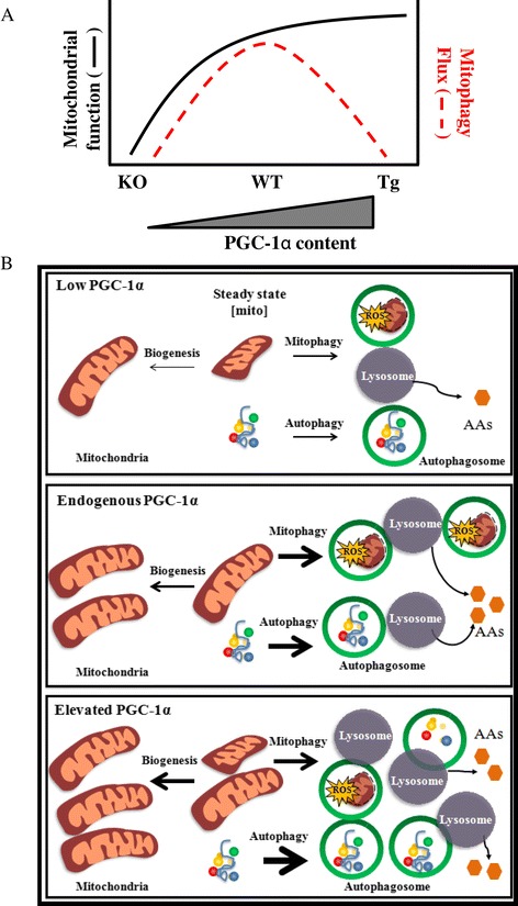
Alterations in skeletal muscle contractile activity necessitate an efficient remodeling mechanism. In particular, mitochondrial turnover is essential for tissue homeostasis during muscle adaptations to chronic use and disuse. While mitochondrial biogenesis appears to be largely governed by the transcriptional co-activator peroxisome proliferator co-activator 1 alpha (PGC-1α), selective mitochondrial autophagy (mitophagy) is thought to mediate organelle degradation. However, whether PGC-1α plays a direct role in autophagy is currently unclear.
To investigate the role of the co-activator in autophagy and mitophagy during skeletal muscle remodeling, PGC-1α knockout (KO) and overexpressing (Tg) animals were unilaterally denervated, a common model of chronic muscle disuse.
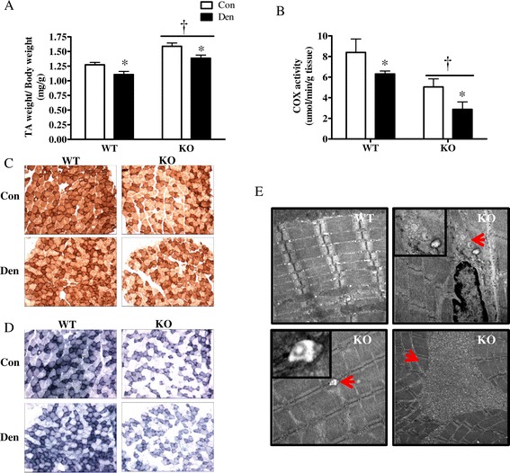
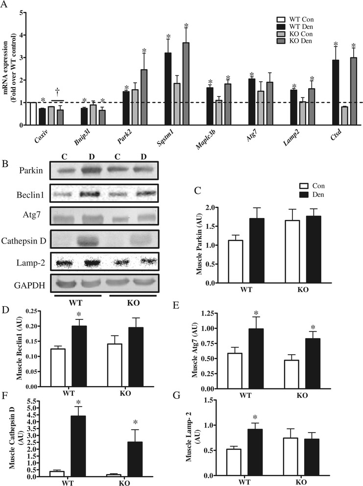
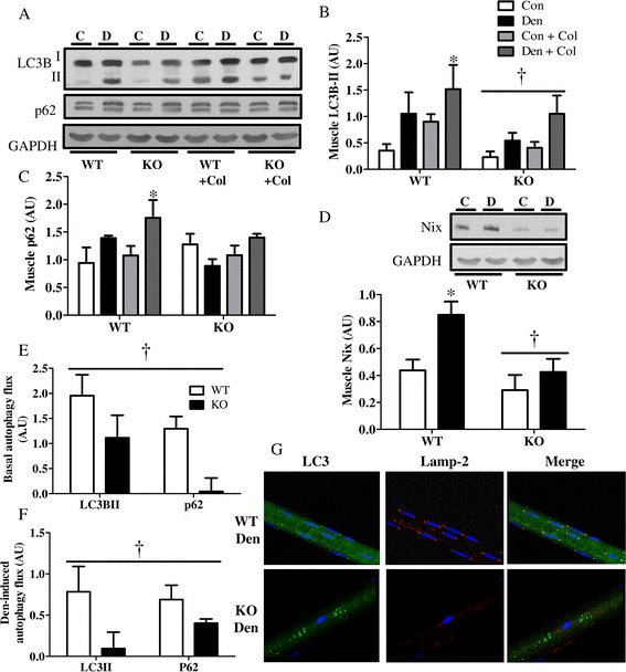
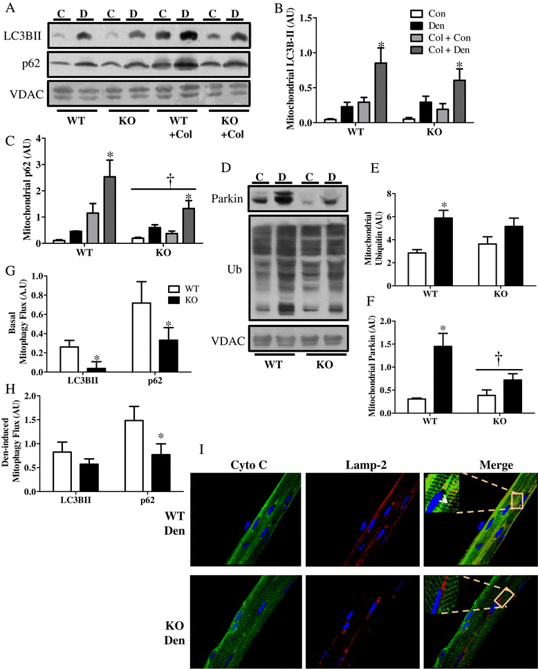
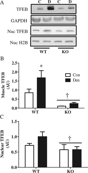
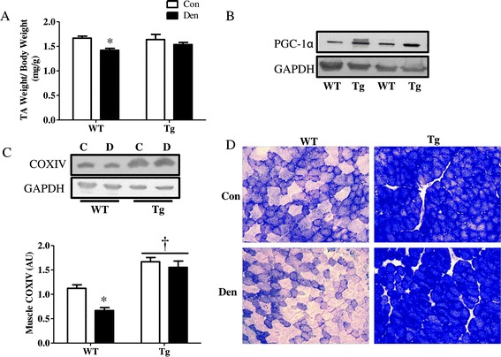
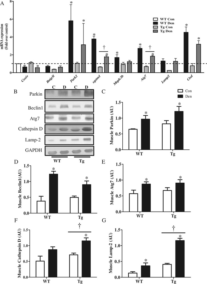
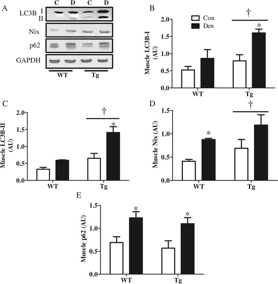
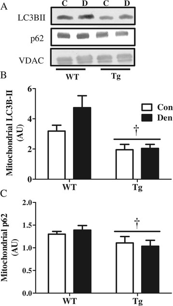
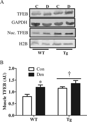
Animals lacking PGC-1α exhibited diminished mitochondrial density alongside myopathic characteristics reminiscent of autophagy-deficient muscle. Denervation promoted an induction in autophagy and lysosomal protein expression in wild-type (WT) animals, which was partially attenuated in KO animals, resulting in reduced autophagy and mitophagy flux. PGC-1α overexpression led to an increase in lysosomal capacity as well as indicators of autophagy flux but exhibited reduced localization of LC3II and p62 to mitochondria, compared to WT animals. A correlation was observed between the levels of the autophagy-lysosome master regulator transcription factor EB (TFEB) and PGC-1α in muscle, supporting their coordinated regulation.
Our investigation has uncovered a regulatory role for PGC-1α in mitochondrial turnover, not only through biogenesis but also via degradation using the autophagy-lysosome machinery. This implies a PGC-1α-mediated cross-talk between these two opposing processes, working to ensure mitochondrial homeostasis during muscle adaptation to chronic disuse.
骨骼肌肉收缩活动的改变需要有效的重塑机制。特别是,线粒体周转对于肌肉适应慢性使用和废用的组织稳态至关重要。虽然线粒体生物发生似乎主要由过氧化物酶体增殖物激活受体共激活因子 1α(PGC-1α)转录共激活因子控制,但选择性线粒体自噬(mitophagy)被认为介导细胞器降解。然而,PGC-1α 是否在自噬中发挥直接作用目前尚不清楚。
为了研究共激活因子在骨骼肌重塑过程中的自噬和线粒体自噬中的作用,对 PGC-1α 敲除(KO)和过表达(Tg)动物进行单侧去神经支配,这是一种常见的慢性肌肉废用模型。
缺乏 PGC-1α 的动物表现出线粒体密度降低,同时伴有类似于自噬缺陷肌肉的肌病特征。去神经支配促进了野生型(WT)动物中自噬和溶酶体蛋白表达的诱导,而 KO 动物中的诱导部分减弱,导致自噬和线粒体自噬通量减少。PGC-1α 过表达导致溶酶体容量增加以及自噬通量的指标增加,但与 WT 动物相比,LC3II 和 p62 在线粒体上的定位减少。观察到肌肉中自噬-溶酶体主调控转录因子 EB(TFEB)和 PGC-1α 之间存在相关性,支持它们的协调调节。
我们的研究揭示了 PGC-1α 在线粒体周转中的调节作用,不仅通过生物发生,而且还通过使用自噬-溶酶体机制进行降解。这意味着 PGC-1α 介导的这两个相反过程之间的交叉对话,有助于确保肌肉适应慢性废用时的线粒体稳态。