Ketschek Andrea, Jones Steven, Spillane Mirela, Korobova Farida, Svitkina Tatyana, Gallo Gianluca
Department of Anatomy and Cell Biology, Shriners Hospitals Pediatric Research Center, Temple University, Philadelphia, Pennsylvania, 19140.
Department of Biology, University of Pennsylvania, Philadelphia, Pennsylvania, 19104.
Dev Neurobiol. 2015 Dec;75(12):1441-61. doi: 10.1002/dneu.22294. Epub 2015 May 27.
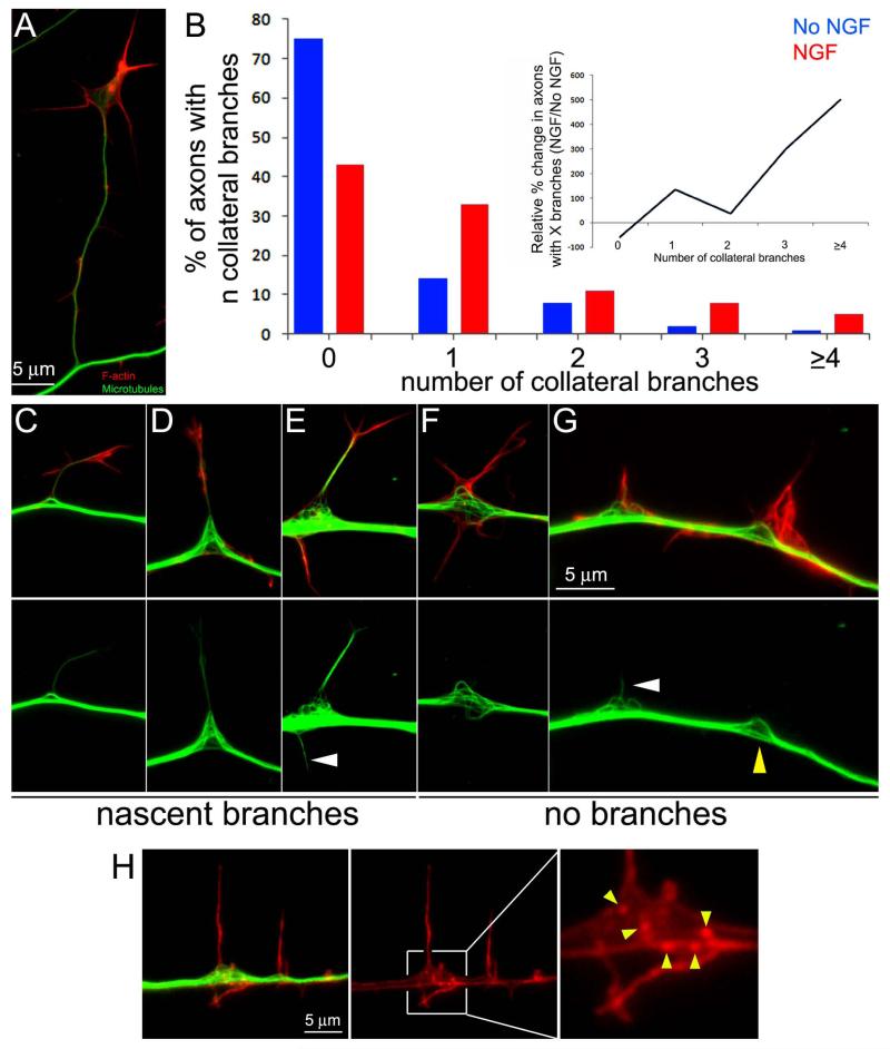
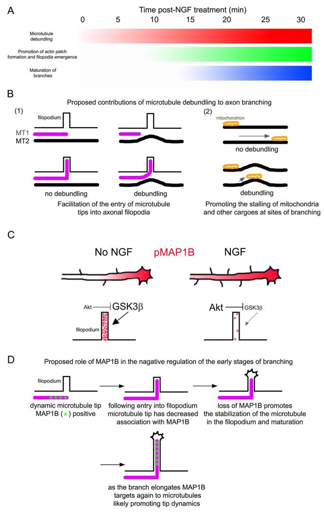
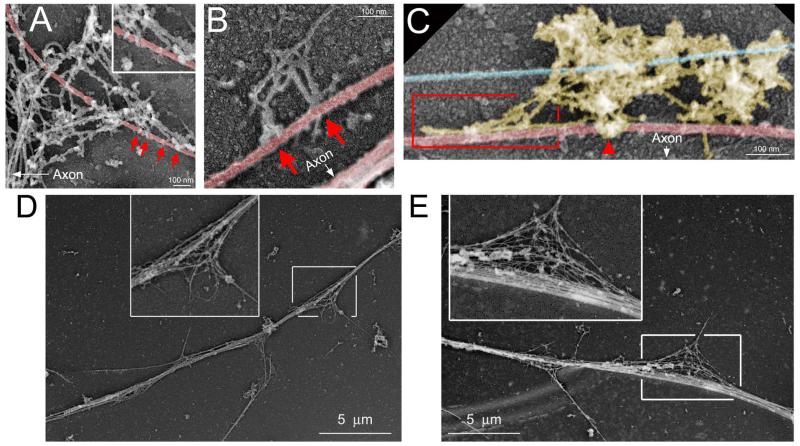
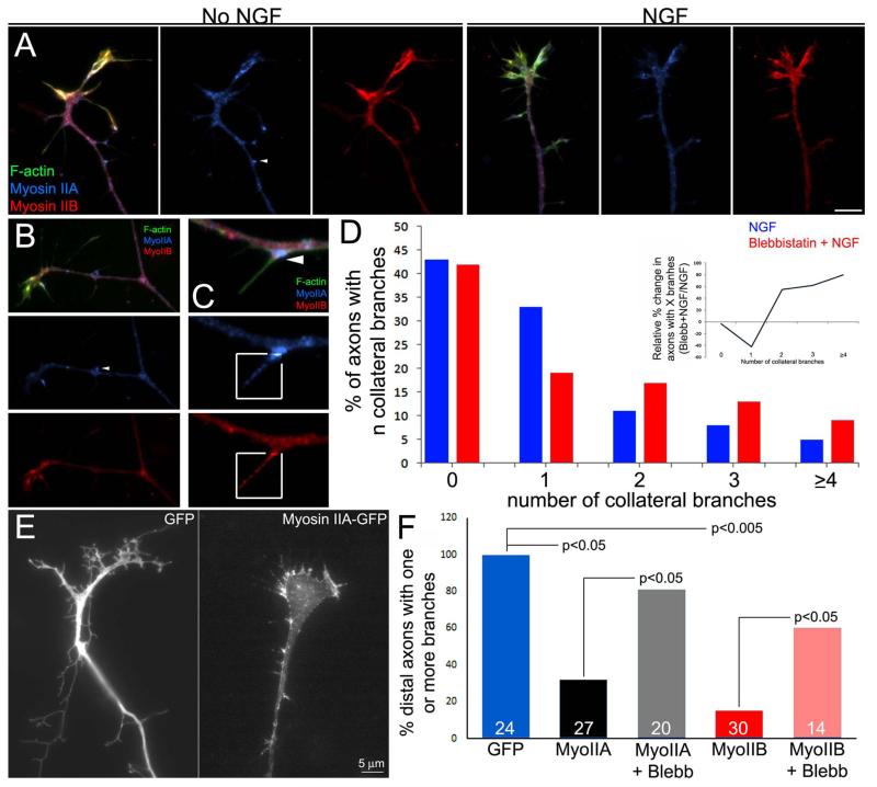
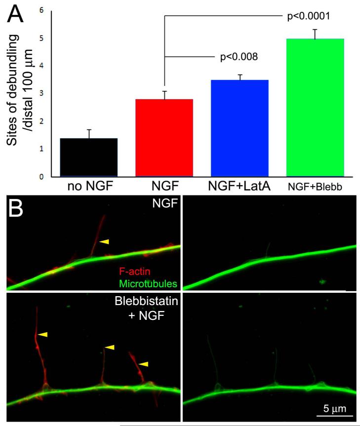
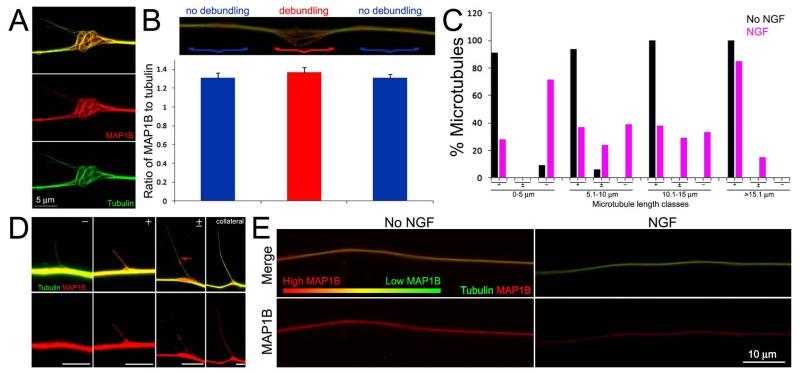
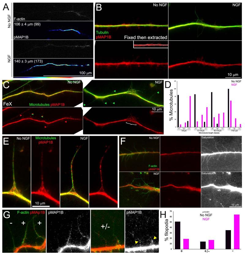
The localized debundling of the axonal microtubule array and the entry of microtubules into axonal filopodia are two defining features of collateral branching. We report that nerve growth factor (NGF), a branch-inducing signal, increases the frequency of microtubule debundling along the axon shaft of chicken embryonic sensory neurons. Sites of debundling correlate strongly with the localized targeting of microtubules into filopodia. Platinum replica electron microscopy suggests physical interactions between debundled microtubules and axonal actin filaments. However, as evidenced by depolymerization of actin filaments and inhibition of myosin II, actomyosin force generation does not promote debundling. In contrast, loss of actin filaments or inhibition of myosin II activity promotes debundling, indicating that axonal actomyosin forces suppress debundling. MAP1B is a microtubule associated protein that represses axon branching. Following treatment with NGF, microtubules penetrating filopodia during the early stages of branching exhibited lower levels of associated MAP1B. NGF increased and decreased the levels of MAP1B phosphorylated at a GSK-3β site (pMAP1B) along the axon shaft and within axonal filopodia, respectively. The levels of MAP1B and pMAP1B were not altered at sites of debundling, relative to the rest of the axon. Unlike the previously determined effects of NGF on the axonal actin cytoskeleton, the effects of NGF on microtubule debundling were not affected by inhibition of protein synthesis. Collectively, these data indicate that NGF promotes localized axonal microtubule debundling, that actomyosin forces antagonize microtubule debundling, and that NGF regulates pMAP1B in axonal filopodia during the early stages of collateral branch formation.
轴突微管阵列的局部解束以及微管进入轴突丝状伪足是侧支分支的两个决定性特征。我们报告称,神经生长因子(NGF)作为一种诱导分支的信号,可增加鸡胚感觉神经元轴突干上微管解束的频率。解束位点与微管向丝状伪足的局部靶向密切相关。铂复型电子显微镜显示解束后的微管与轴突肌动蛋白丝之间存在物理相互作用。然而,肌动蛋白丝的解聚和肌球蛋白II的抑制表明,肌动球蛋白产生的力并不促进解束。相反,肌动蛋白丝的缺失或肌球蛋白II活性的抑制会促进解束,这表明轴突肌动球蛋白力会抑制解束。微管相关蛋白1B(MAP1B)是一种抑制轴突分支的微管相关蛋白。用NGF处理后,在分支早期穿透丝状伪足的微管上,MAP1B的相关水平较低。NGF分别增加和降低了轴突干和轴突丝状伪足内GSK-3β位点磷酸化的MAP1B(pMAP1B)水平。相对于轴突的其他部位,解束位点处的MAP1B和pMAP1B水平没有改变。与之前确定的NGF对轴突肌动蛋白细胞骨架的作用不同,NGF对微管解束的作用不受蛋白质合成抑制的影响。总的来说,这些数据表明,NGF促进轴突微管的局部解束,肌动球蛋白力拮抗微管解束,并且NGF在侧支分支形成的早期阶段调节轴突丝状伪足中的pMAP1B。