• 文献检索
  • 文档翻译
  • 深度研究
  • 学术资讯
  • Suppr Zotero 插件Zotero 插件
  • 邀请有礼
  • 套餐&价格
  • 历史记录
应用&插件
Suppr Zotero 插件Zotero 插件浏览器插件Mac 客户端Windows 客户端微信小程序
定价
高级版会员购买积分包购买API积分包
服务
文献检索文档翻译深度研究API 文档MCP 服务
关于我们
关于 Suppr公司介绍联系我们用户协议隐私条款
关注我们

Suppr 超能文献

核心技术专利:CN118964589B侵权必究
粤ICP备2023148730 号-1Suppr @ 2026

文献检索

告别复杂PubMed语法,用中文像聊天一样搜索,搜遍4000万医学文献。AI智能推荐,让科研检索更轻松。

立即免费搜索

文件翻译

保留排版,准确专业,支持PDF/Word/PPT等文件格式,支持 12+语言互译。

免费翻译文档

深度研究

AI帮你快速写综述,25分钟生成高质量综述,智能提取关键信息,辅助科研写作。

立即免费体验

Optomap超广角成像可识别糖尿病视网膜病变患者的其他视网膜异常。

Optomap ultrawide field imaging identifies additional retinal abnormalities in patients with diabetic retinopathy.

作者信息

Price Liam D, Au Stephanie, Chong N Victor

机构信息

Oxford Eye Hospital, University of Oxford, Oxford, UK.

University of Hong Kong, Hong Kong SAR, People's Republic of China.

出版信息

Clin Ophthalmol. 2015 Mar 24;9:527-31. doi: 10.2147/OPTH.S79448. eCollection 2015.

DOI:10.2147/OPTH.S79448
PMID:25848202
原文链接:https://pmc.ncbi.nlm.nih.gov/articles/PMC4376301/
Abstract

PURPOSE

To compare diabetic retinopathy (DR) severity grading between Optomap ultrawide field scanning laser ophthalmoscope (UWFSLO) 200° images and an Early Treatment Diabetic Retinopathy Study (ETDRS) seven-standard field view.

METHODS

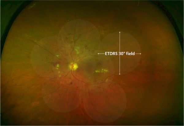
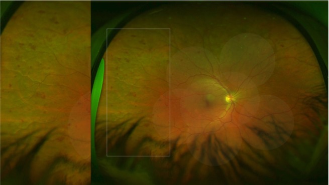
Optomap UWFSLO images (total: 266) were retrospectively selected for evidence of DR from a database of eye clinic attendees. The Optomap UWFSLO images were graded for DR severity by two masked assessors. An ETDRS seven-field mask was overlaid on the Optomap UWFSLO images, and the DR grade was assessed for the region inside the mask. Any interassessor discrepancies were adjudicated by a senior retinal specialist. Kappa agreement levels were used for statistical analysis.

RESULTS

Fifty images (19%) (P<0.001) were assigned a higher DR level in the Optomap UWFSLO view compared to the ETDRS seven-field view, which resulted in 40 images (15%) (P<0.001) receiving a higher DR severity grade. DR severity grades in the ETDRS seven-field view compared with the Optomap UWFSLO view were identical in 85% (226) of the images and within one severity level in 100% (266) of the images. Agreement between the two views was substantial: unweighted κ was 0.74±0.04 (95% confidence interval: 0.67-0.81) and weighted κ was 0.80±0.03 (95% confidence interval: 0.74-0.86).

CONCLUSION

Compared to the ETDRS seven-field view, a significant minority of patients are diagnosed with more severe DR when using the Optomap UWFSLO view. The clinical significance of additional peripheral lesions requires evaluation in future prospective studies using large cohorts.

摘要

目的

比较Optomap超广角扫描激光检眼镜(UWFSLO)200°图像与糖尿病视网膜病变早期治疗研究(ETDRS)七标准视野下糖尿病视网膜病变(DR)的严重程度分级。

方法

从眼科门诊患者数据库中回顾性选取Optomap UWFSLO图像(共266张)以寻找DR证据。由两名盲法评估者对Optomap UWFSLO图像的DR严重程度进行分级。将ETDRS七视野模板覆盖在Optomap UWFSLO图像上,并对模板内区域的DR分级进行评估。评估者之间的任何差异由一位资深视网膜专家进行判定。采用Kappa一致性水平进行统计分析。

结果

与ETDRS七视野相比,50张图像(19%)(P<0.001)在Optomap UWFSLO视野下被判定为更高的DR水平,这使得40张图像(15%)(P<0.001)获得更高的DR严重程度分级。ETDRS七视野与Optomap UWFSLO视野下的DR严重程度分级在85%(226张)的图像中相同,在100%(266张)的图像中相差不超过一个严重程度级别。两种视野之间的一致性较高:未加权κ值为0.74±0.04(95%置信区间:[0.67, 0.81]),加权κ值为0.80±0.03(95%置信区间:[0.74, 0.86])。

结论

与ETDRS七视野相比,使用Optomap UWFSLO视野时,有相当少数的患者被诊断为更严重的DR。额外周边病变的临床意义需要在未来使用大型队列的前瞻性研究中进行评估。

https://cdn.ncbi.nlm.nih.gov/pmc/blobs/45d8/4376301/fa174aa5a865/opth-9-527Fig2.jpg
https://cdn.ncbi.nlm.nih.gov/pmc/blobs/45d8/4376301/763437a649c7/opth-9-527Fig1.jpg
https://cdn.ncbi.nlm.nih.gov/pmc/blobs/45d8/4376301/fa174aa5a865/opth-9-527Fig2.jpg
https://cdn.ncbi.nlm.nih.gov/pmc/blobs/45d8/4376301/763437a649c7/opth-9-527Fig1.jpg
https://cdn.ncbi.nlm.nih.gov/pmc/blobs/45d8/4376301/fa174aa5a865/opth-9-527Fig2.jpg

相似文献

1
Optomap ultrawide field imaging identifies additional retinal abnormalities in patients with diabetic retinopathy.Optomap超广角成像可识别糖尿病视网膜病变患者的其他视网膜异常。
Clin Ophthalmol. 2015 Mar 24;9:527-31. doi: 10.2147/OPTH.S79448. eCollection 2015.
2
Nonmydriatic ultrawide field retinal imaging compared with dilated standard 7-field 35-mm photography and retinal specialist examination for evaluation of diabetic retinopathy.非散瞳超广角视网膜成像与散瞳标准 7 区域 35mm 摄影及视网膜专科医师检查在糖尿病视网膜病变评估中的比较。
Am J Ophthalmol. 2012 Sep;154(3):549-559.e2. doi: 10.1016/j.ajo.2012.03.019. Epub 2012 May 23.
3
Assessment of diabetic retinopathy using nonmydriatic ultra-widefield scanning laser ophthalmoscopy (Optomap) compared with ETDRS 7-field stereo photography.应用非散瞳超广角激光扫描检眼镜(Optomap)评估糖尿病性视网膜病变与 ETDRS 7 区域立体摄影比较。
Diabetes Care. 2012 Dec;35(12):2459-63. doi: 10.2337/dc12-0346. Epub 2012 Aug 21.
4
[Diabetic retinopathy: comparison of the diagnostic features of ultra-widefield scanning laser ophthalmoscopy Optomap with ETDRS 7-field fundus photography].[糖尿病视网膜病变:超广角扫描激光检眼镜Optomap与ETDRS七视野眼底摄影诊断特征的比较]
Ophthalmologe. 2011 Feb;108(2):117-23. doi: 10.1007/s00347-010-2226-4.
5
Peripheral lesions identified by mydriatic ultrawide field imaging: distribution and potential impact on diabetic retinopathy severity.散瞳超广角成像识别的周边病变:分布及对糖尿病视网膜病变严重程度的潜在影响。
Ophthalmology. 2013 Dec;120(12):2587-2595. doi: 10.1016/j.ophtha.2013.05.004. Epub 2013 Jun 15.
6
Comparison of Widefield Laser Ophthalmoscopy and ETDRS Retinal Area for Diabetic Retinopathy.广角激光眼底镜检查与糖尿病视网膜病变早期治疗糖尿病性视网膜病变研究(ETDRS)视网膜区域的比较
Ophthalmol Sci. 2022 Jun 28;2(4):100190. doi: 10.1016/j.xops.2022.100190. eCollection 2022 Dec.
7
Comparison of Early Treatment Diabetic Retinopathy Study Standard 7-Field Imaging With Ultrawide-Field Imaging for Determining Severity of Diabetic Retinopathy.比较早期糖尿病性视网膜病变研究标准 7 字段成像与超广角成像用于确定糖尿病性视网膜病变的严重程度。
JAMA Ophthalmol. 2019 Jan 1;137(1):65-73. doi: 10.1001/jamaophthalmol.2018.4982.
8
Hemorrhage and/or Microaneurysm Severity and Count in Ultrawide Field Images and Early Treatment Diabetic Retinopathy Study Photography.超广角图像中的出血和/或微动脉瘤严重程度和数量与早期糖尿病性视网膜病变研究摄影。
Ophthalmology. 2017 Jul;124(7):970-976. doi: 10.1016/j.ophtha.2017.02.012. Epub 2017 Mar 20.
9
Automated Retinal Imaging System (ARIS) compared with ETDRS protocol color stereoscopic retinal photography to assess level of diabetic retinopathy.自动化视网膜成像系统(ARIS)与 ETDRS 协议彩色立体视网膜摄影比较,评估糖尿病视网膜病变的程度。
Diabetes Technol Ther. 2012 Jun;14(6):515-22. doi: 10.1089/dia.2011.0270. Epub 2012 Mar 2.
10
Comparison of Optomap ultrawide-field imaging versus slit-lamp biomicroscopy for assessment of diabetic retinopathy in a real-life clinic.在现实临床环境中,Optomap超广角成像与裂隙灯生物显微镜检查在糖尿病视网膜病变评估中的比较。
Clin Ophthalmol. 2014 Jul 29;8:1413-7. doi: 10.2147/OPTH.S66700. eCollection 2014.

引用本文的文献

1
Quantitative ultra-widefield fluorescein angiography biomarkers in diabetic retinopathy and association with treatment and progression.糖尿病视网膜病变中的定量超广角荧光素血管造影生物标志物及其与治疗和病情进展的关联
Clin Ophthalmol. 2024 Dec 27;18:4019-4028. doi: 10.2147/OPTH.S472071. eCollection 2024.
2
Quantitative Biomarkers of Diabetic Retinopathy Using Ultra-Widefield Fluorescein Angiography.利用超广角荧光素血管造影术检测糖尿病视网膜病变的定量生物标志物
Clin Ophthalmol. 2024 Jul 8;18:1961-1970. doi: 10.2147/OPTH.S462223. eCollection 2024.
3
Peripheral retinal lesions in diabetic retinopathy on ultra-widefield imaging.

本文引用的文献

1
Peripheral lesions identified by mydriatic ultrawide field imaging: distribution and potential impact on diabetic retinopathy severity.散瞳超广角成像识别的周边病变:分布及对糖尿病视网膜病变严重程度的潜在影响。
Ophthalmology. 2013 Dec;120(12):2587-2595. doi: 10.1016/j.ophtha.2013.05.004. Epub 2013 Jun 15.
2
Ultra-wide field retinal imaging in detection, classification, and management of diabetic retinopathy.超广角视网膜成像在糖尿病视网膜病变的检测、分类及管理中的应用
Semin Ophthalmol. 2012 Sep-Nov;27(5-6):221-7. doi: 10.3109/08820538.2012.708812.
3
Assessment of diabetic retinopathy using nonmydriatic ultra-widefield scanning laser ophthalmoscopy (Optomap) compared with ETDRS 7-field stereo photography.
超广角成像下糖尿病视网膜病变的周边视网膜病变
Saudi J Ophthalmol. 2024 Jan 6;38(2):123-131. doi: 10.4103/sjopt.sjopt_151_23. eCollection 2024 Apr-Jun.
4
Improving diabetic retinopathy screening at the point of care: integrating telemedicine to overcome current challenges.改善即时护理中的糖尿病视网膜病变筛查:整合远程医疗以克服当前挑战。
BMC Ophthalmol. 2024 Jun 14;24(1):256. doi: 10.1186/s12886-024-03508-4.
5
Assessing Diabetic Retinopathy Staging With AI: A Comparative Analysis Between Pseudocolor and LED Imaging.评估糖尿病视网膜病变分期的人工智能技术:伪彩与 LED 成像的对比分析。
Transl Vis Sci Technol. 2024 Mar 1;13(3):11. doi: 10.1167/tvst.13.3.11.
6
Macula vs periphery in diabetic retinopathy: OCT-angiography and ultrawide field fluorescein angiography imaging of retinal non perfusion.糖尿病视网膜病变中的黄斑与周边:视网膜无灌注的 OCT-angiography 和超广角荧光素血管造影成像。
Eye (Lond). 2024 Jun;38(9):1668-1673. doi: 10.1038/s41433-024-02989-3. Epub 2024 Feb 24.
7
Assessment of area and structural irregularity of retinal layers in diabetic retinopathy using machine learning and image processing techniques.利用机器学习和图像处理技术评估糖尿病视网膜病变的视网膜层面积和结构不规则性。
Sci Rep. 2024 Feb 18;14(1):4013. doi: 10.1038/s41598-024-54535-6.
8
Advantages of the Utilization of Wide-Field OCT and Wide-Field OCT Angiography in Clinical Practice.临床实践中使用广角光学相干断层扫描(OCT)和广角OCT血管造影的优势。
Diagnostics (Basel). 2024 Feb 1;14(3):321. doi: 10.3390/diagnostics14030321.
9
Imaging Modalities for Assessing the Vascular Component of Diabetic Retinal Disease: Review and Consensus for an Updated Staging System.评估糖尿病视网膜病变血管成分的成像方式:更新分期系统的综述与共识
Ophthalmol Sci. 2023 Dec 10;4(3):100449. doi: 10.1016/j.xops.2023.100449. eCollection 2024 May-Jun.
10
Wide-field imaging with smartphone based fundus camera: grading of severity of diabetic retinopathy and locating peripheral lesions in diabetic retinopathy.基于智能手机的眼底相机广角成像:糖尿病视网膜病变严重程度分级和糖尿病视网膜病变周边病变定位。
Eye (Lond). 2024 Jun;38(8):1471-1476. doi: 10.1038/s41433-024-02928-2. Epub 2024 Jan 31.
应用非散瞳超广角激光扫描检眼镜(Optomap)评估糖尿病性视网膜病变与 ETDRS 7 区域立体摄影比较。
Diabetes Care. 2012 Dec;35(12):2459-63. doi: 10.2337/dc12-0346. Epub 2012 Aug 21.
4
Nonmydriatic ultrawide field retinal imaging compared with dilated standard 7-field 35-mm photography and retinal specialist examination for evaluation of diabetic retinopathy.非散瞳超广角视网膜成像与散瞳标准 7 区域 35mm 摄影及视网膜专科医师检查在糖尿病视网膜病变评估中的比较。
Am J Ophthalmol. 2012 Sep;154(3):549-559.e2. doi: 10.1016/j.ajo.2012.03.019. Epub 2012 May 23.
5
Peripheral retinal ischaemia, as evaluated by ultra-widefield fluorescein angiography, is associated with diabetic macular oedema.周边视网膜缺血,通过超广角荧光素血管造影评估,与糖尿病黄斑水肿有关。
Br J Ophthalmol. 2012 May;96(5):694-8. doi: 10.1136/bjophthalmol-2011-300774. Epub 2012 Mar 15.
6
Comparison of film and digital fundus photographs in eyes of individuals with diabetes mellitus.糖尿病患者眼底的数码和传统摄影照片比较。
Invest Ophthalmol Vis Sci. 2011 Aug 3;52(9):6168-73. doi: 10.1167/iovs.11-7321.
7
Comparability of digital photography with the ETDRS film protocol for evaluation of diabetic retinopathy severity.数字摄影与 ETDRS 眼底照相协议评估糖尿病视网膜病变严重程度的比较。
Invest Ophthalmol Vis Sci. 2011 Jul 1;52(7):4717-25. doi: 10.1167/iovs.10-6303.
8
Differential diameter responses in macular and peripheral retinal arterioles may contribute to the regional distribution of diabetic retinopathy lesions.黄斑和周边视网膜小动脉的直径差异反应可能有助于糖尿病视网膜病变病变的区域性分布。
Graefes Arch Clin Exp Ophthalmol. 2011 Mar;249(3):407-12. doi: 10.1007/s00417-010-1549-9. Epub 2010 Nov 11.
9
Peripheral vessel leakage (PVL): a new angiographic finding in diabetic retinopathy identified with ultra wide-field fluorescein angiography.外周血管渗漏(PVL):糖尿病视网膜病变中通过超广角荧光素血管造影术发现的一种新的血管造影表现。
Semin Ophthalmol. 2010 Jan-Mar;25(1-2):27-33. doi: 10.3109/08820538.2010.481239.
10
Screening for diabetic retinopathy: a comparative trial of photography and scanning laser ophthalmoscopy.糖尿病性视网膜病变的筛查:摄影术与扫描激光检眼镜的对比研究。
Ophthalmologica. 2010;224(4):251-7. doi: 10.1159/000284351. Epub 2010 Feb 10.