Samsel Leigh, McCoy J Philip
National Heart, Lung, and Blood Institute, NIH, Bethesda, MD 20892, United States.
J Immunol Methods. 2015 Aug;423:52-9. doi: 10.1016/j.jim.2015.03.019. Epub 2015 Apr 7.
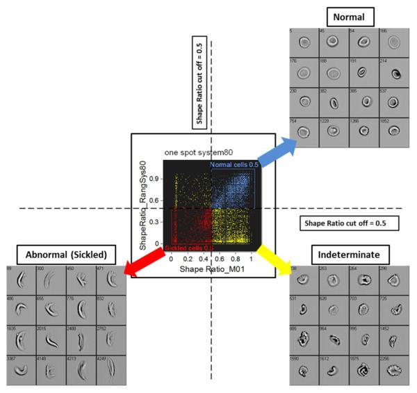
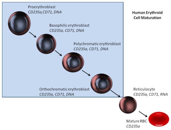
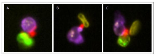
Erythroid cell maturation and diseases affecting erythrocytes are frequently accompanied by morphologic and immunophenotypic changes to these cells. In the past, these changes have been assessed primarily through the use of manual microscopy, which substantially limits the statistical rigor, throughput, and objectivity of these studies. Imaging flow cytometry provides a technology to examine both the morphology of cells as well as to quantify the staining intensity and signal distribution of numerous fluorescent markers on a cell-by-cell basis with high throughput in a statistically robust manner, and thus is ideally suited to studying erythroid cell biology. To date imaging flow cytometry has been used to study erythrocytes in three areas: 1) erythroid cell maturation, 2) sickle cell disease, and 3) infectious diseases such as malaria. In the maturation studies, imaging flow cytometry can closely recapitulate known stages of maturation and has led to the identification of a new population of erythroid cell precursors. In sickle cell disease, imaging flow cytometry provides a robust method to quantify sickled erythrocytes and to identify cellular aggregates linked to morbidities, and in malaria, imaging flow cytometry has been used to screen for new chemotherapeutic agents. These studies have demonstrated the value of imaging flow cytometry for investigations of erythrocyte biology and pathology.
红系细胞成熟以及影响红细胞的疾病常常伴随着这些细胞的形态学和免疫表型变化。过去,这些变化主要通过手工显微镜检查来评估,这在很大程度上限制了这些研究的统计严谨性、通量和客观性。成像流式细胞术提供了一种技术,能够以高通量、统计稳健的方式逐个细胞地检查细胞形态以及量化众多荧光标记物的染色强度和信号分布,因此非常适合研究红系细胞生物学。迄今为止,成像流式细胞术已用于三个领域研究红细胞:1)红系细胞成熟,2)镰状细胞病,3)疟疾等传染病。在成熟研究中,成像流式细胞术可以精确重现已知的成熟阶段,并已导致鉴定出一种新的红系细胞前体群体。在镰状细胞病中,成像流式细胞术提供了一种强大的方法来量化镰状红细胞并识别与发病机制相关的细胞聚集体,而在疟疾研究中,成像流式细胞术已用于筛选新的化疗药物。这些研究证明了成像流式细胞术在红细胞生物学和病理学研究中的价值。