Rijlaarsdam Martin A, Tax David M J, Gillis Ad J M, Dorssers Lambert C J, Koestler Devin C, de Ridder Jeroen, Looijenga Leendert H J
Department of Pathology, Erasmus MC Cancer Institute-University Medical Center Rotterdam, Rotterdam, The Netherlands.
Faculty of Electrical Engineering, Mathematics and Computer Science Intelligent Systems-Delft Bioinformatics Lab, Technical University of Delft, Delft, The Netherlands.
PLoS One. 2015 Apr 10;10(4):e0122146. doi: 10.1371/journal.pone.0122146. eCollection 2015.
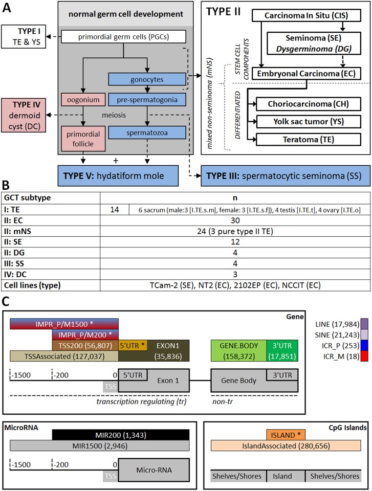
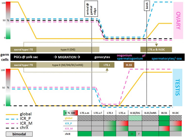
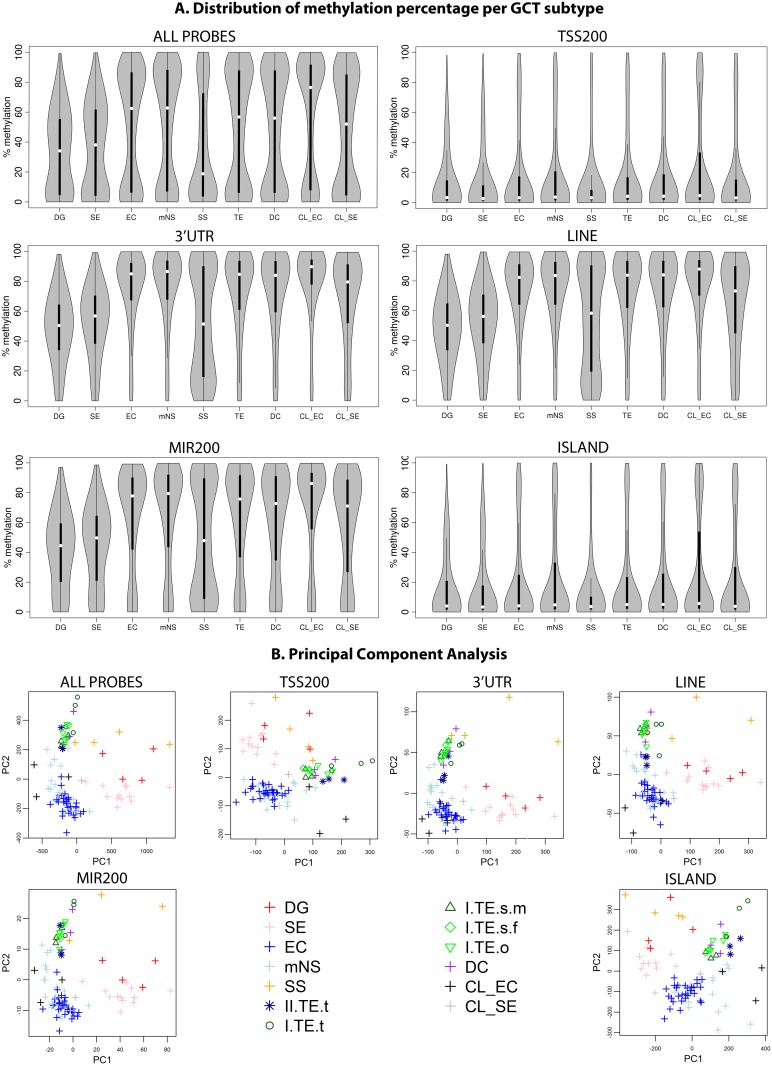
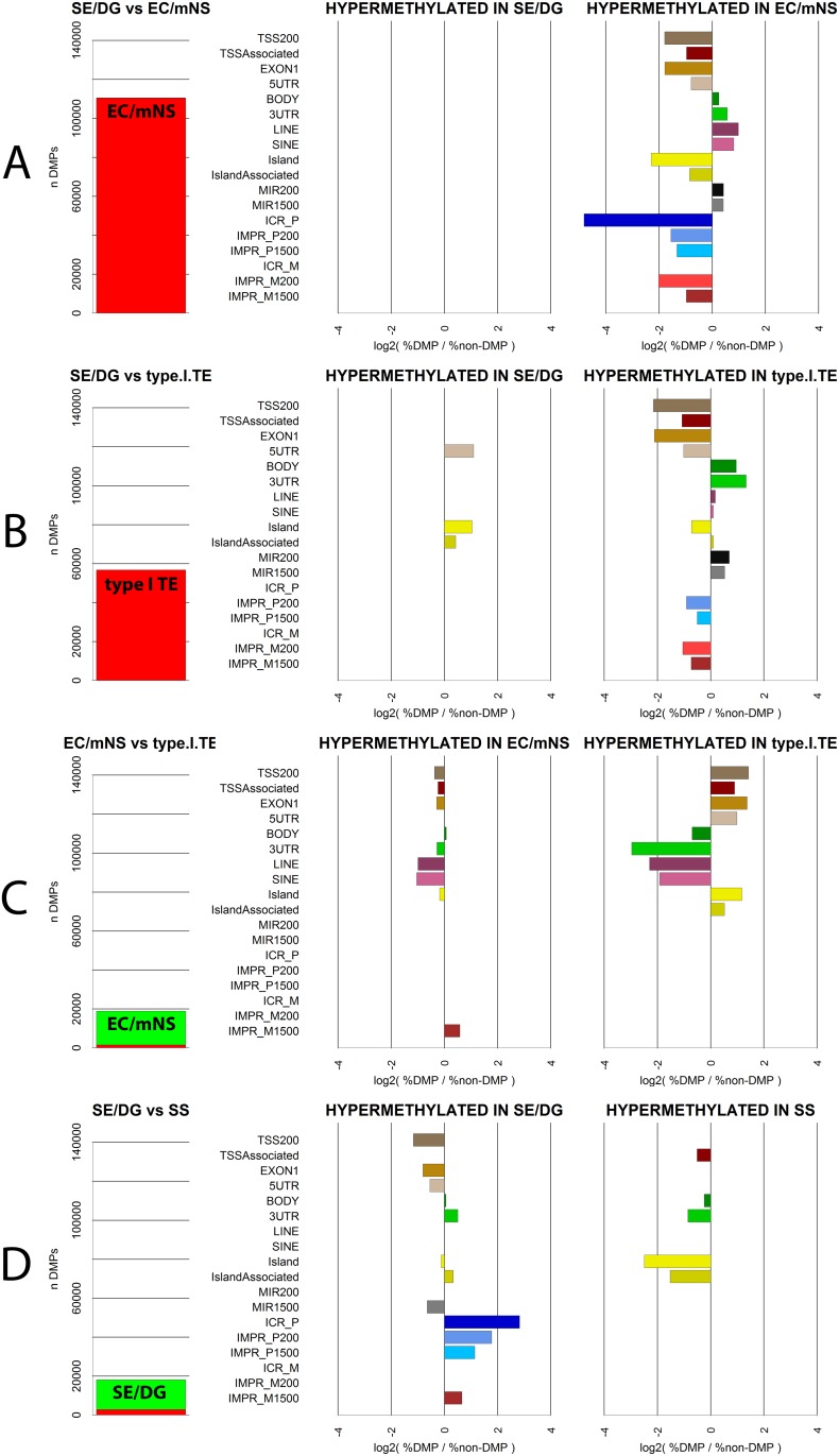
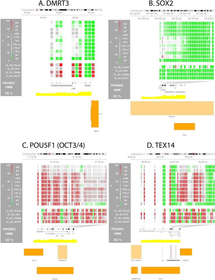
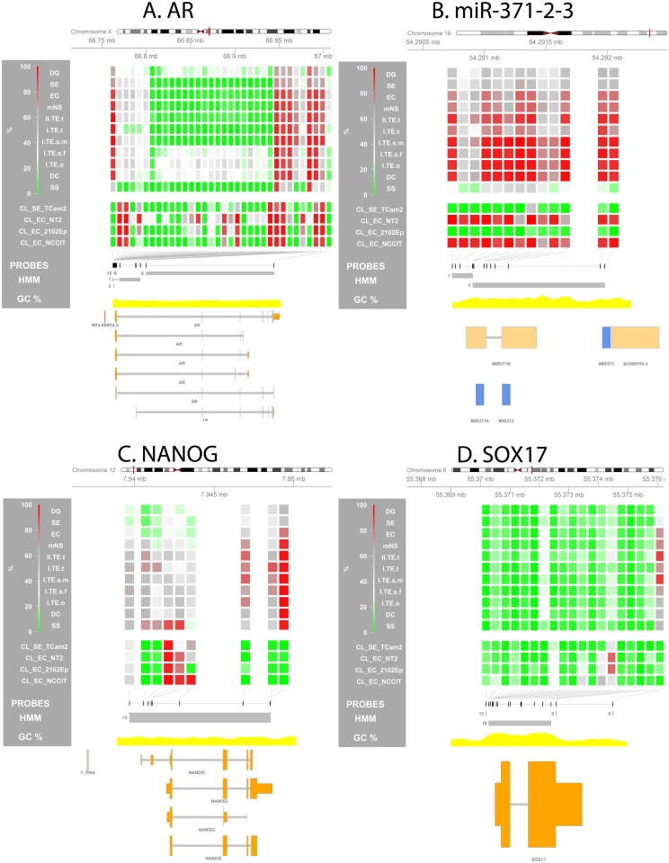
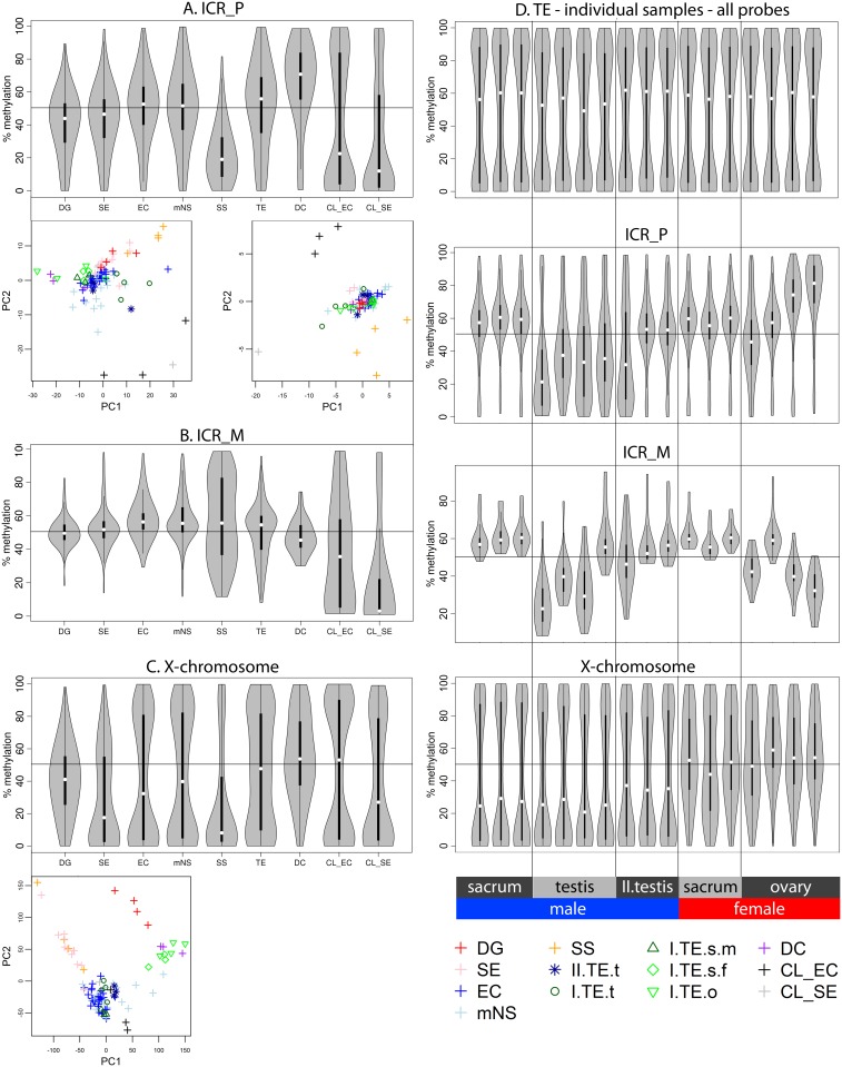
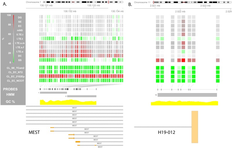
The cell of origin of the five subtypes (I-V) of germ cell tumors (GCTs) are assumed to be germ cells from different maturation stages. This is (potentially) reflected in their methylation status as fetal maturing primordial germ cells are globally demethylated during migration from the yolk sac to the gonad. Imprinted regions are erased in the gonad and later become uniparentally imprinted according to fetal sex. Here, 91 GCTs (type I-IV) and four cell lines were profiled (Illumina's HumanMethylation450BeadChip). Data was pre-processed controlling for cross hybridization, SNPs, detection rate, probe-type bias and batch effects. The annotation was extended, covering snRNAs/microRNAs, repeat elements and imprinted regions. A Hidden Markov Model-based genome segmentation was devised to identify differentially methylated genomic regions. Methylation profiles allowed for separation of clusters of non-seminomas (type II), seminomas/dysgerminomas (type II), spermatocytic seminomas (type III) and teratomas/dermoid cysts (type I/IV). The seminomas, dysgerminomas and spermatocytic seminomas were globally hypomethylated, in line with previous reports and their demethylated precursor. Differential methylation and imprinting status between subtypes reflected their presumed cell of origin. Ovarian type I teratomas and dermoid cysts showed (partial) sex specific uniparental maternal imprinting. The spermatocytic seminomas showed uniparental paternal imprinting while testicular teratomas exhibited partial imprinting erasure. Somatic imprinting in type II GCTs might indicate a cell of origin after global demethylation but before imprinting erasure. This is earlier than previously described, but agrees with the totipotent/embryonic stem cell like potential of type II GCTs and their rare extra-gonadal localization. The results support the common origin of the type I teratomas and show strong similarity between ovarian type I teratomas and dermoid cysts. In conclusion, we identified specific and global methylation differences between GCT subtypes, providing insight into their developmental timing and underlying developmental biology. Data and extended annotation are deposited at GEO (GSE58538 and GPL18809).
生殖细胞肿瘤(GCT)的五种亚型(I - V)的起源细胞被认为是来自不同成熟阶段的生殖细胞。这一点(可能)反映在它们的甲基化状态上,因为胎儿成熟的原始生殖细胞在从卵黄囊迁移到性腺的过程中会发生全基因组去甲基化。印记区域在性腺中被擦除,随后根据胎儿性别变成单亲印记。在此,对91个GCT(I - IV型)和四个细胞系进行了分析(Illumina公司的HumanMethylation450BeadChip)。对数据进行了预处理,以控制交叉杂交、单核苷酸多态性(SNP)、检测率、探针类型偏差和批次效应。注释范围得到了扩展,涵盖了小核仁RNA/微小RNA、重复元件和印记区域。设计了一种基于隐马尔可夫模型的基因组分割方法来识别差异甲基化的基因组区域。甲基化谱允许区分非精原细胞瘤(II型)、精原细胞瘤/无性细胞瘤(II型)、精母细胞性精原细胞瘤(III型)和畸胎瘤/皮样囊肿(I/IV型)的簇。精原细胞瘤、无性细胞瘤和精母细胞性精原细胞瘤整体上是低甲基化的,这与之前的报道及其去甲基化的前体一致。各亚型之间的差异甲基化和印记状态反映了它们假定的起源细胞。卵巢I型畸胎瘤和皮样囊肿表现出(部分)性别特异性的单亲母系印记。精母细胞性精原细胞瘤表现出单亲父系印记,而睾丸畸胎瘤表现出部分印记擦除。II型GCT中的体细胞印记可能表明起源细胞处于全基因组去甲基化之后但印记擦除之前的阶段。这比之前描述的时间更早,但与II型GCT的全能/胚胎干细胞样潜能及其罕见的性腺外定位情况相符。这些结果支持了I型畸胎瘤的共同起源,并显示出卵巢I型畸胎瘤和皮样囊肿之间有很强的相似性。总之,我们确定了GCT亚型之间特定的和整体的甲基化差异,为它们的发育时间和潜在的发育生物学提供了见解。数据和扩展注释已存入基因表达综合数据库(GEO)(GSE58538和GPL18809)。