Molecular Design and Chemical Biology, F. Hoffmann-La Roche Ltd , Grenzacherstrasse 124, 4070 Basel, Switzerland.
Instituto de Biología Molecular de Barcelona (IBMB), CSIC , Barcelona Science Park, Baldiri Reixach 15, 08028 Barcelona, Spain.
IUCrJ. 2015 Jan 27;2(Pt 2):177-87. doi: 10.1107/S2052252515000238. eCollection 2015 Mar 1.
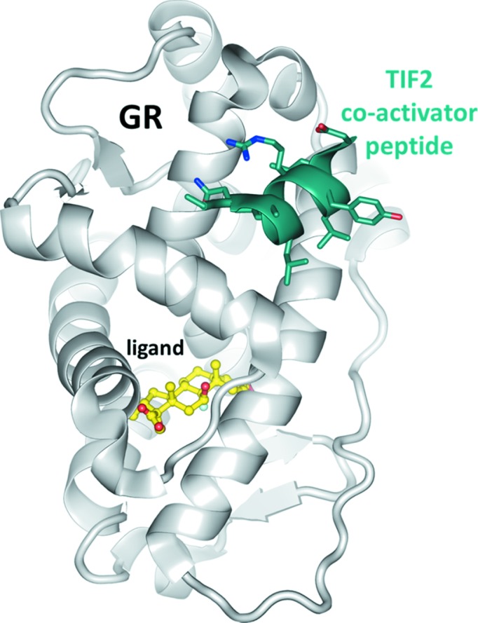
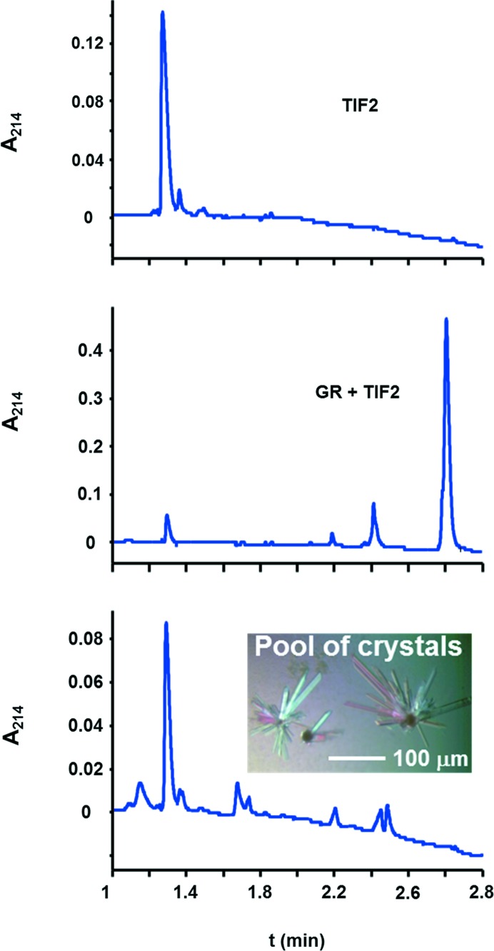
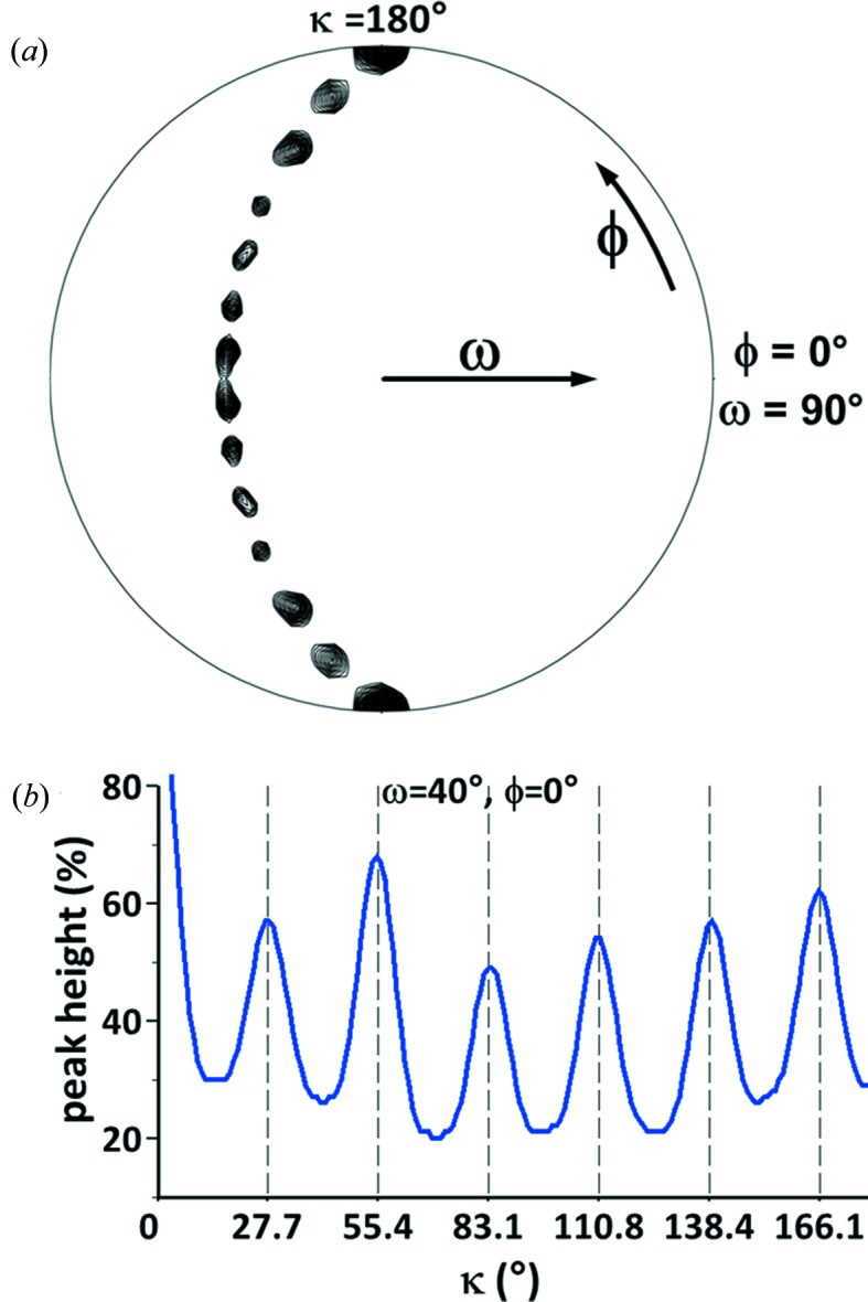
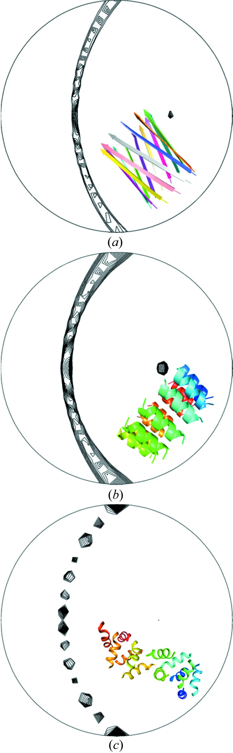
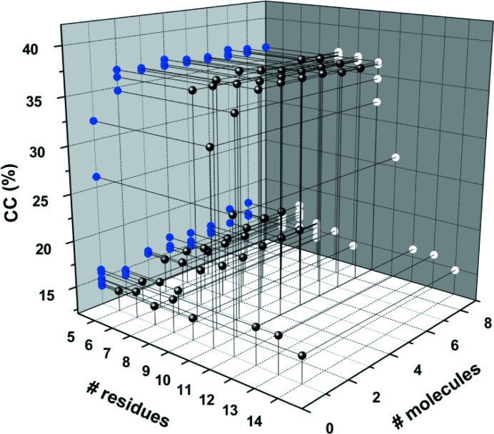
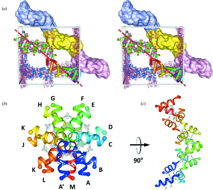
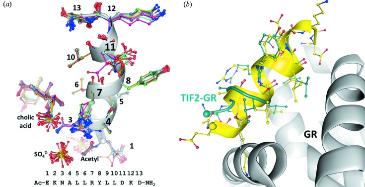
Nuclear hormone receptors are cytoplasm-based transcription factors that bind a ligand, translate to the nucleus and initiate gene transcription in complex with a co-activator such as TIF2 (transcriptional intermediary factor 2). For structural studies the co-activator is usually mimicked by a peptide of circa 13 residues, which for the largest part forms an α-helix when bound to the receptor. The aim was to co-crystallize the glucocorticoid receptor in complex with a ligand and the TIF2 co-activator peptide. The 1.82 Å resolution diffraction data obtained from the crystal could not be phased by molecular replacement using the known receptor structures. HPLC analysis of the crystals revealed the absence of the receptor and indicated that only the co-activator peptide was present. The self-rotation function displayed 13-fold rotational symmetry, which initiated an exhaustive but unsuccessful molecular-replacement approach using motifs of 13-fold symmetry such as α- and β-barrels in various geometries. The structure was ultimately determined by using a single α-helix and the software ARCIMBOLDO, which assembles fragments placed by PHASER before using them as seeds for density modification model building in SHELXE. Systematic variation of the helix length revealed upper and lower size limits for successful structure determination. A beautiful but unanticipated structure was obtained that forms superhelices with left-handed twist throughout the crystal, stabilized by ligand interactions. Together with the increasing diversity of structural elements in the Protein Data Bank the results from TIF2 confirm the potential of fragment-based molecular replacement to significantly accelerate the phasing step for native diffraction data at around 2 Å resolution.
核激素受体是位于细胞质中的转录因子,它们与配体结合,转位到细胞核内,并与转录中介因子 2(TIF2)等共激活因子形成复合物,启动基因转录。对于结构研究,共激活因子通常由大约 13 个残基的肽模拟,当与受体结合时,该肽大部分形成α-螺旋。本研究的目的是共结晶糖皮质激素受体与配体和 TIF2 共激活肽复合物。从晶体中获得的 1.82 Å分辨率衍射数据无法通过使用已知受体结构的分子置换进行相位推断。晶体的 HPLC 分析显示没有受体,表明只有共激活肽存在。自旋转功能显示出 13 重旋转对称性,这引发了一种详尽但不成功的分子置换方法,使用了各种几何形状的 13 重对称基序,如α-和β-桶。最终,使用单个α-螺旋和软件 ARCIMBOLDO 确定了结构,该软件在使用 PHASER 放置的片段作为 SHELXE 中密度修饰模型构建的种子之前,将这些片段组装在一起。螺旋长度的系统变化揭示了成功结构确定的上限和下限尺寸。得到了一个美丽但出乎意料的结构,该结构在整个晶体中形成左手扭曲的超螺旋,由配体相互作用稳定。与蛋白质数据库中结构元件的多样性增加一起,TIF2 的结果证实了基于片段的分子置换在大约 2 Å 分辨率的天然衍射数据的相确定步骤中具有显著加速的潜力。