• 文献检索
  • 文档翻译
  • 深度研究
  • 学术资讯
  • Suppr Zotero 插件Zotero 插件
  • 邀请有礼
  • 套餐&价格
  • 历史记录
应用&插件
Suppr Zotero 插件Zotero 插件浏览器插件Mac 客户端Windows 客户端微信小程序
定价
高级版会员购买积分包购买API积分包
服务
文献检索文档翻译深度研究API 文档MCP 服务
关于我们
关于 Suppr公司介绍联系我们用户协议隐私条款
关注我们

Suppr 超能文献

核心技术专利:CN118964589B侵权必究
粤ICP备2023148730 号-1Suppr @ 2026

文献检索

告别复杂PubMed语法,用中文像聊天一样搜索,搜遍4000万医学文献。AI智能推荐,让科研检索更轻松。

立即免费搜索

文件翻译

保留排版,准确专业,支持PDF/Word/PPT等文件格式,支持 12+语言互译。

免费翻译文档

深度研究

AI帮你快速写综述,25分钟生成高质量综述,智能提取关键信息,辅助科研写作。

立即免费体验

可视化骨髓间充质干细胞移植修复脊髓损伤

Visual bone marrow mesenchymal stem cell transplantation in the repair of spinal cord injury.

作者信息

Zhang Rui-Ping, Xu Cheng, Liu Yin, Li Jian-Ding, Xie Jun

机构信息

Department of Radiology, First Hospital, Shanxi Medical University, Taiyuan, Shanxi Province, China ; Molecular Imaging Program at Stanford, Stanford University, Stanford, CA, USA.

Department of Radiology, Shanxi Provincial People's Hospital, Taiyuan, Shanxi Province, China.

出版信息

Neural Regen Res. 2015 Mar;10(3):404-11. doi: 10.4103/1673-5374.153688.

DOI:10.4103/1673-5374.153688
PMID:25878588
原文链接:https://pmc.ncbi.nlm.nih.gov/articles/PMC4396102/
Abstract

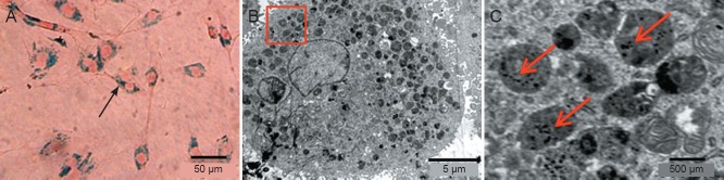
An important factor in improving functional recovery from spinal cord injury using stem cells is maximizing the number of transplanted cells at the lesion site. Here, we established a contusion model of spinal cord injury by dropping a weight onto the spinal cord at T7-8. Superparamagnetic iron oxide-labeled bone marrow mesenchymal stem cells were transplanted into the injured spinal cord via the subarachnoid space. An outer magnetic field was used to successfully guide the labeled cells to the lesion site. Prussian blue staining showed that more bone marrow mesenchymal stem cells reached the lesion site in these rats than in those without magnetic guidance or superparamagnetic iron oxide labeling, and immunofluorescence revealed a greater number of complete axons at the lesion site. Moreover, the Basso, Beattie and Bresnahan (BBB) locomotor rating scale scores were the highest in rats with superparamagnetic labeling and magnetic guidance. Our data confirm that superparamagnetic iron oxide nanoparticles effectively label bone marrow mesenchymal stem cells and impart sufficient magnetism to respond to the external magnetic field guides. More importantly, superparamagnetic iron oxide-labeled bone marrow mesenchymal stem cells can be dynamically and non-invasively tracked in vivo using magnetic resonance imaging. Superparamagnetic iron oxide labeling of bone marrow mesenchymal stem cells coupled with magnetic guidance offers a promising avenue for the clinical treatment of spinal cord injury.

摘要

利用干细胞改善脊髓损伤功能恢复的一个重要因素是使损伤部位移植细胞数量最大化。在此,我们通过在T7 - 8节段向脊髓上掉落重物建立了脊髓损伤的挫伤模型。将超顺磁性氧化铁标记的骨髓间充质干细胞经蛛网膜下腔移植到损伤的脊髓中。使用外部磁场成功地将标记细胞引导至损伤部位。普鲁士蓝染色显示,与未进行磁引导或超顺磁性氧化铁标记的大鼠相比,这些大鼠中有更多的骨髓间充质干细胞到达损伤部位,免疫荧光显示损伤部位有更多完整的轴突。此外,在具有超顺磁性标记和磁引导的大鼠中,Basso、Beattie和Bresnahan(BBB)运动评分量表得分最高。我们的数据证实,超顺磁性氧化铁纳米颗粒能有效标记骨髓间充质干细胞,并赋予足够的磁性以响应外部磁场引导。更重要的是,使用磁共振成像可以在体内动态、无创地追踪超顺磁性氧化铁标记的骨髓间充质干细胞。骨髓间充质干细胞的超顺磁性氧化铁标记与磁引导相结合为脊髓损伤的临床治疗提供了一条有前景的途径。

https://cdn.ncbi.nlm.nih.gov/pmc/blobs/bf76/4396102/8e248335c831/NRR-10-404-g006.jpg
https://cdn.ncbi.nlm.nih.gov/pmc/blobs/bf76/4396102/6b2645937a03/NRR-10-404-g001.jpg
https://cdn.ncbi.nlm.nih.gov/pmc/blobs/bf76/4396102/0702ce5abcd4/NRR-10-404-g002.jpg
https://cdn.ncbi.nlm.nih.gov/pmc/blobs/bf76/4396102/d4977b235344/NRR-10-404-g003.jpg
https://cdn.ncbi.nlm.nih.gov/pmc/blobs/bf76/4396102/d8eaadb0ba79/NRR-10-404-g004.jpg
https://cdn.ncbi.nlm.nih.gov/pmc/blobs/bf76/4396102/5e13b75ac8b7/NRR-10-404-g005.jpg
https://cdn.ncbi.nlm.nih.gov/pmc/blobs/bf76/4396102/8e248335c831/NRR-10-404-g006.jpg

相似文献

1
Visual bone marrow mesenchymal stem cell transplantation in the repair of spinal cord injury.可视化骨髓间充质干细胞移植修复脊髓损伤
Neural Regen Res. 2015 Mar;10(3):404-11. doi: 10.4103/1673-5374.153688.
2
Chronic spinal cord injury treated with transplanted autologous bone marrow-derived mesenchymal stem cells tracked by magnetic resonance imaging: a case report.磁共振成像追踪自体骨髓间充质干细胞移植治疗慢性脊髓损伤:一例报告
J Med Case Rep. 2015 Apr 9;9:79. doi: 10.1186/s13256-015-0535-6.
3
In vivo tracking of neuronal-like cells by magnetic resonance in rabbit models of spinal cord injury.在脊髓损伤兔模型中通过磁共振对类神经元细胞进行体内示踪。
Neural Regen Res. 2013 Dec 25;8(36):3373-81. doi: 10.3969/j.issn.1673-5374.2013.36.002.
4
In vivo tracking of stem cells in brain and spinal cord injury.脑和脊髓损伤中干细胞的体内追踪
Prog Brain Res. 2007;161:367-83. doi: 10.1016/S0079-6123(06)61026-1.
5
Bone marrow stem cells and polymer hydrogels--two strategies for spinal cord injury repair.骨髓干细胞与聚合物水凝胶——脊髓损伤修复的两种策略。
Cell Mol Neurobiol. 2006 Oct-Nov;26(7-8):1113-29. doi: 10.1007/s10571-006-9007-2. Epub 2006 Apr 22.
6
Magnetic resonance tracking of transplanted bone marrow and embryonic stem cells labeled by iron oxide nanoparticles in rat brain and spinal cord.大鼠脑和脊髓中氧化铁纳米颗粒标记的移植骨髓和胚胎干细胞的磁共振追踪
J Neurosci Res. 2004 Apr 15;76(2):232-43. doi: 10.1002/jnr.20041.
7
Noggin Protein can Induce the Differentiation of Rat Bone Marrow Mesenchymal Stem Cells to Neurons and Repair Spinal Cord Injury.Noggin 蛋白可诱导大鼠骨髓间充质干细胞向神经元分化并修复脊髓损伤。
Discov Med. 2023 Dec;35(179):956-964. doi: 10.24976/Discov.Med.202335179.91.
8
Effects of combination treatment with transcranial magnetic stimulation and bone marrow mesenchymal stem cell transplantation or Raf inhibition on spinal cord injury in rats.经颅磁刺激联合骨髓间充质干细胞移植或 Raf 抑制对大鼠脊髓损伤的影响。
Mol Med Rep. 2021 Apr;23(4). doi: 10.3892/mmr.2021.11934. Epub 2021 Mar 2.
9
Electrophysiological functional recovery in a rat model of spinal cord hemisection injury following bone marrow-derived mesenchymal stem cell transplantation under hypothermia.低温下骨髓间充质干细胞移植后脊髓半切损伤大鼠模型的电生理功能恢复
Neural Regen Res. 2012 Apr 5;7(10):749-55. doi: 10.3969/j.issn.1673-5374.2012.10.005.
10
Effect of External Magnetic Field on the Efficacy of ADSC Transplantation in Rats with Spinal Cord Injury.磁场对大鼠脊髓损伤后脂肪间充质干细胞移植疗效的影响。
Turk Neurosurg. 2024;34(5):888-897. doi: 10.5137/1019-5149.JTN.45113-23.2.

引用本文的文献

1
Prussian blue nanotechnology in the treatment of spinal cord injury: application and challenges.普鲁士蓝纳米技术在脊髓损伤治疗中的应用与挑战
Front Bioeng Biotechnol. 2024 Sep 11;12:1474711. doi: 10.3389/fbioe.2024.1474711. eCollection 2024.
2
Iron Oxide Nanoparticles in Regenerative Medicine and Tissue Engineering.再生医学与组织工程中的氧化铁纳米颗粒
Nanomaterials (Basel). 2021 Sep 8;11(9):2337. doi: 10.3390/nano11092337.
3
Stem Cell Therapy for Spinal Cord Injury.脊髓损伤的干细胞治疗。

本文引用的文献

1
In vivo tracking of neuronal-like cells by magnetic resonance in rabbit models of spinal cord injury.在脊髓损伤兔模型中通过磁共振对类神经元细胞进行体内示踪。
Neural Regen Res. 2013 Dec 25;8(36):3373-81. doi: 10.3969/j.issn.1673-5374.2013.36.002.
2
Cultivation and identification of rat bone marrow-derived mesenchymal stem cells.大鼠骨髓间充质干细胞的培养与鉴定
Mol Med Rep. 2014 Aug;10(2):755-60. doi: 10.3892/mmr.2014.2264. Epub 2014 May 22.
3
Transplantation of neurotrophin-3-expressing bone mesenchymal stem cells improves recovery in a rat model of spinal cord injury.
Cell Transplant. 2021 Jan-Dec;30:963689721989266. doi: 10.1177/0963689721989266.
4
Progress in Stem Cell Therapy for Spinal Cord Injury.脊髓损伤的干细胞治疗进展
Stem Cells Int. 2020 Nov 5;2020:2853650. doi: 10.1155/2020/2853650. eCollection 2020.
5
Magnetic Composite Biomaterials for Neural Regeneration.用于神经再生的磁性复合生物材料
Front Bioeng Biotechnol. 2019 Jul 25;7:179. doi: 10.3389/fbioe.2019.00179. eCollection 2019.
6
Stem Cells and Labeling for Spinal Cord Injury.干细胞与脊髓损伤的标记
Int J Mol Sci. 2016 Dec 26;18(1):6. doi: 10.3390/ijms18010006.
7
Increased Understanding of Stem Cell Behavior in Neurodegenerative and Neuromuscular Disorders by Use of Noninvasive Cell Imaging.通过使用非侵入性细胞成像技术增进对神经退行性疾病和神经肌肉疾病中干细胞行为的理解。
Stem Cells Int. 2016;2016:6235687. doi: 10.1155/2016/6235687. Epub 2016 Feb 22.
8
Protective effect of bone marrow mesenchymal stem cells on PC12 cells apoptosis mediated by TAG1.骨髓间充质干细胞对TAG1介导的PC12细胞凋亡的保护作用
Int J Clin Exp Pathol. 2015 Oct 1;8(10):12093-100. eCollection 2015.
表达神经营养因子-3的骨髓间充质干细胞移植可改善大鼠脊髓损伤模型的恢复情况。
Acta Neurochir (Wien). 2014 Jul;156(7):1409-18. doi: 10.1007/s00701-014-2089-6. Epub 2014 Apr 18.
4
The isolation and cultivation of bone marrow stem cells and evaluation of differences for neural-like cells differentiation under the induction with neurotrophic factors.骨髓干细胞的分离培养及其在神经营养因子诱导下向神经样细胞分化差异的评价。
Cytotechnology. 2014 Dec;66(6):1007-19. doi: 10.1007/s10616-013-9654-3. Epub 2014 Jan 1.
5
Comparison of functional and histological outcomes after intralesional, intracisternal, and intravenous transplantation of human bone marrow-derived mesenchymal stromal cells in a rat model of spinal cord injury.在大鼠脊髓损伤模型中,比较经皮内、蛛网膜下腔和静脉内移植人骨髓间充质基质细胞后的功能和组织学结果。
Acta Neurochir (Wien). 2013 Oct;155(10):1943-50. doi: 10.1007/s00701-013-1799-5. Epub 2013 Jul 3.
6
Effect of neuroglobin genetically modified bone marrow mesenchymal stem cells transplantation on spinal cord injury in rabbits.神经球蛋白基因修饰骨髓间充质干细胞移植对兔脊髓损伤的影响。
PLoS One. 2013 May 2;8(5):e63444. doi: 10.1371/journal.pone.0063444. Print 2013.
7
In vivo magnetic resonance imaging of hyperpolarized silicon particles.体内磁共振成像的超极化硅颗粒。
Nat Nanotechnol. 2013 May;8(5):363-8. doi: 10.1038/nnano.2013.65. Epub 2013 May 5.
8
Recent advances in superparamagnetic iron oxide nanoparticles for cellular imaging and targeted therapy research.超顺磁性氧化铁纳米粒子在细胞成像和靶向治疗研究中的最新进展。
Curr Pharm Des. 2013;19(37):6575-93. doi: 10.2174/1381612811319370003.
9
In vivo magnetic resonance imaging tracking of C6 glioma cells labeled with superparamagnetic iron oxide nanoparticles.用超顺磁性氧化铁纳米颗粒标记的C6胶质瘤细胞的体内磁共振成像追踪
Einstein (Sao Paulo). 2012 Apr-Jun;10(2):164-70. doi: 10.1590/s1679-45082012000200009.
10
MR characterization of focal nodular hyperplasia: gadoxetic acid versus superparamagnetic iron oxide imaging.MRI 对肝局灶性结节增生的特征分析:钆塞酸二钠对比超顺磁性氧化铁成像。
Magn Reson Med Sci. 2012;11(3):163-9. doi: 10.2463/mrms.11.163.