Zou Wei, Chen Qiu-Xin, Sun Xiao-Wei, Chi Qing-Bin, Kuang Hong-Yu, Yu Xue-Ping, Dai Xiao-Hong
Third Department of Acupuncture, First Affiliated Hospital, Heilongjiang University of Chinese Medicine, Harbin, Heilongjiang Province, China.
Heilongjiang University of Chinese Medicine, Harbin, Heilongjiang Province, China.
Neural Regen Res. 2015 Mar;10(3):457-62. doi: 10.4103/1673-5374.153696.
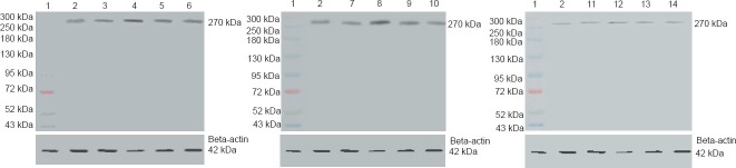
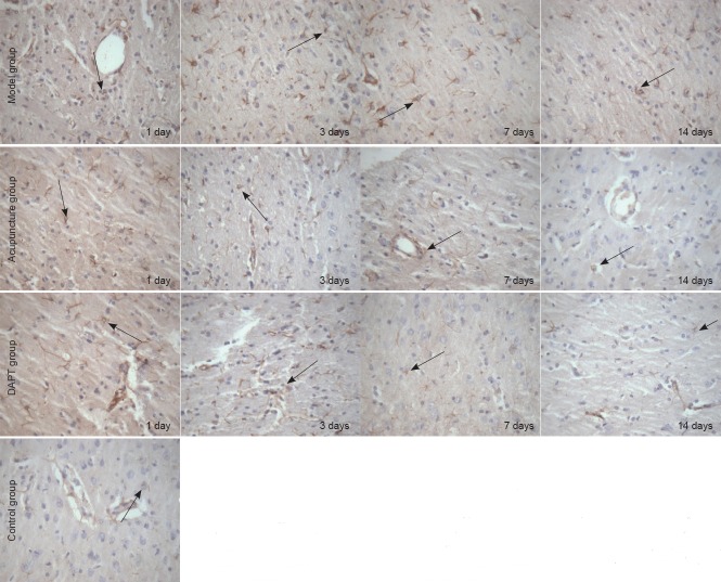
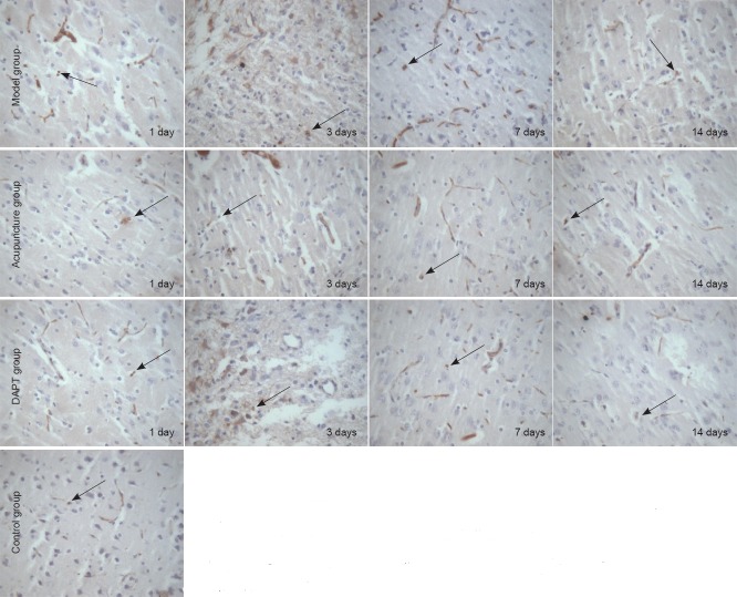
Notch pathway activation maintains neural stem cells in a proliferating state and increases nerve repair capacity. To date, studies have rarely focused on changes or damage to signal transduction pathways during cerebral hemorrhage. Here, we examined the effect of acupuncture in a rat model of cerebral hemorrhage. We examined four groups: in the control group, rats received no treatment. In the model group, cerebral hemorrhage models were established by infusing non-heparinized blood into the brain. In the acupuncture group, modeled rats had Baihui (DU20) and Qubin (GB7) acupoints treated once a day for 30 minutes. In the DAPT group, modeled rats had 0.15 μg/mL DAPT solution (10 mL) infused into the brain. Immunohistochemistry and western blot results showed that acupuncture effectively inhibits Notch1 and Hes1 protein expression in rat basal ganglia. These inhibitory effects were identical to DAPT, a Notch signaling pathway inhibitor. Our results suggest that acupuncture has a neuroprotective effect on cerebral hemorrhage by inhibiting Notch-Hes signaling pathway transduction in rat basal ganglia after cerebral hemorrhage.
Notch信号通路激活可使神经干细胞维持在增殖状态并增强神经修复能力。迄今为止,针对脑出血期间信号转导通路的变化或损伤的研究很少。在此,我们研究了针刺对大鼠脑出血模型的影响。我们设置了四组:对照组大鼠不接受任何治疗。模型组通过向脑内注入非肝素化血液建立脑出血模型。针刺组对造模大鼠的百会(DU20)和曲鬓(GB7)穴位每天治疗1次,每次30分钟。DAPT组对造模大鼠脑内注入0.15μg/mL DAPT溶液(10 mL)。免疫组织化学和蛋白质印迹结果显示,针刺可有效抑制大鼠基底神经节中Notch1和Hes1蛋白表达。这些抑制作用与Notch信号通路抑制剂DAPT的作用相同。我们的结果表明,针刺通过抑制脑出血后大鼠基底神经节中的Notch-Hes信号通路转导,对脑出血具有神经保护作用。