Shahidi Shoaleh, Zadeh Nahal Kazerooni, Sharafeddin Farahnaz, Shahab Shahriar, Bahrampour Ehsan, Hamedani Shahram
Department of Oral and Maxillofacial Radiology, Shiraz University of Medical Sciences, Shiraz, Iran.
Postgraduate Student, Department of Oral and Maxillofacial Radiology, Shiraz University of Medical Sciences, Shiraz, Iran.
Dent Res J (Isfahan). 2015 Mar-Apr;12(2):161-6.
This study was aimed to compare the diagnostic accuracy and feasibility of cone beam computed tomography (CBCT) with phosphor storage plate (PSP) in detection of simulated occlusal secondary caries.
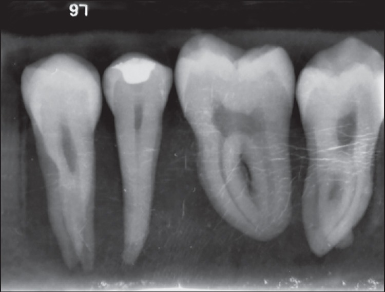
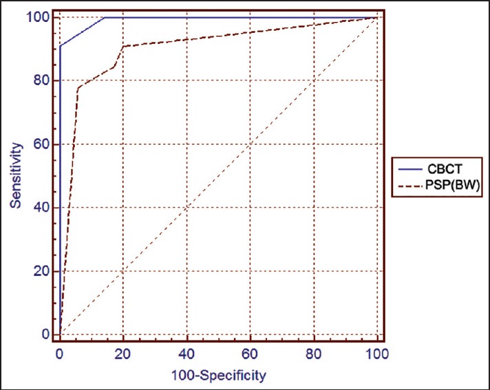
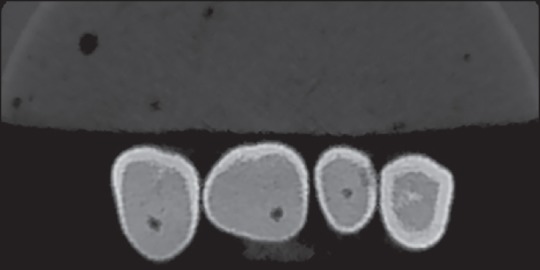
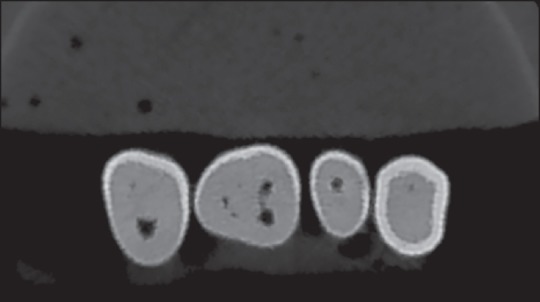
In this in vitro descriptive-comparative study, a total of 80 slots of class I cavities were prepared on 80 extracted human premolars. Then, 40 teeth were randomly selected out of this sample and artificial carious lesions were created on these teeth by a round diamond bur no. 1/2. All 80 teeth were restored with amalgam fillings and radiographs were taken, both with PSP system and CBCT. All images were evaluated by three calibrated observers. The area under the receiver operating characteristic curve was used to compare the diagnostic accuracy of two systems. SPSS (SPSS Inc., Chicago, IL, USA) was adopted for statistical analysis. The difference between Az value of bitewing and CBCT methods were compared by pairwise comparison method. The inter- and intra-operator agreement was assessed by kappa analysis (P < 0.05).
The mean Az value for bitewings and CBCT was 0.903 and 0.994, respectively. Significant differences were found between PSP and CBCT (P = 0.010). The kappa value for inter-observer agreement was 0.68 and 0.76 for PSP and CBCT, respectively. The kappa value for intra-observer agreement was 0.698 (observer 1, P = 0.000), 0.766 (observer 2, P = 0.000) and 0.716 (observer 3, P = 0.000) in PSP method, and 0.816 (observer 1, P = 0.000), 0.653 (observer 2, P = 0.000) and 0.744 (observer 3, P = 0.000) in CBCT method.
This in vitro study, with a limited number of samples, showed that the New Tom VGI Flex CBCT system was more accurate than the PSP in detecting the simulated small secondary occlusal caries under amalgam restoration.
本研究旨在比较锥形束计算机断层扫描(CBCT)与磷光存储板(PSP)在检测模拟咬合继发龋方面的诊断准确性和可行性。
在这项体外描述性比较研究中,在80颗拔除的人类前磨牙上制备了总共80个I类洞。然后,从该样本中随机选择40颗牙齿,用1/2号圆钻在这些牙齿上制造人工龋损。所有80颗牙齿均用银汞合金充填并拍摄X线片,包括使用PSP系统和CBCT。所有图像由三名经过校准的观察者进行评估。采用受试者工作特征曲线下面积比较两种系统的诊断准确性。采用SPSS(SPSS公司,美国伊利诺伊州芝加哥)进行统计分析。采用两两比较法比较咬合翼片和CBCT方法的Az值差异。通过kappa分析评估观察者间和观察者内的一致性(P<0.05)。
咬合翼片和CBCT的平均Az值分别为0.903和0.994。PSP和CBCT之间存在显著差异(P=0.010)。PSP和CBCT观察者间一致性的kappa值分别为0.68和0.76。PSP方法中观察者内一致性的kappa值分别为0.698(观察者1,P=0.000)、0.766(观察者2,P=0.000)和0.716(观察者3,P=0.000),CBCT方法中分别为0.816(观察者1,P=0.000)、0.653(观察者2,P=0.000)和0.744(观察者3,P=0.000)。
这项样本数量有限的体外研究表明,New Tom VGI Flex CBCT系统在检测银汞合金修复下模拟的小咬合继发龋方面比PSP更准确。