• 文献检索
  • 文档翻译
  • 深度研究
  • 学术资讯
  • Suppr Zotero 插件Zotero 插件
  • 邀请有礼
  • 套餐&价格
  • 历史记录
应用&插件
Suppr Zotero 插件Zotero 插件浏览器插件Mac 客户端Windows 客户端微信小程序
定价
高级版会员购买积分包购买API积分包
服务
文献检索文档翻译深度研究API 文档MCP 服务
关于我们
关于 Suppr公司介绍联系我们用户协议隐私条款
关注我们

Suppr 超能文献

核心技术专利:CN118964589B侵权必究
粤ICP备2023148730 号-1Suppr @ 2026

文献检索

告别复杂PubMed语法,用中文像聊天一样搜索,搜遍4000万医学文献。AI智能推荐,让科研检索更轻松。

立即免费搜索

文件翻译

保留排版,准确专业,支持PDF/Word/PPT等文件格式,支持 12+语言互译。

免费翻译文档

深度研究

AI帮你快速写综述,25分钟生成高质量综述,智能提取关键信息,辅助科研写作。

立即免费体验

通过评分系统鉴别脂肪瘤和非典型脂肪瘤性肿瘤:血管增多对脂肪肉瘤发病机制的影响

Differentiation of lipoma and atypical lipomatous tumor by a scoring system: implication of increased vascularity on pathogenesis of liposarcoma.

作者信息

Nagano Satoshi, Yokouchi Masahiro, Setoguchi Takao, Ishidou Yasuhiro, Sasaki Hiromi, Shimada Hirofumi, Komiya Setsuro

机构信息

Department of Orthopaedic Surgery, Graduate School of Medical and Dental Sciences, Kagoshima University, 8-35-1 Sakuragaoka, Kagoshima-city, Kagoshima, 890-8520, Japan.

The Near-Future Locomotor Organ Medicine Creation Course (Kusunoki Kai), Graduate School of Medical and Dental Sciences, Kagoshima University, Kagoshima, Japan.

出版信息

BMC Musculoskelet Disord. 2015 Feb 22;16:36. doi: 10.1186/s12891-015-0491-8.

DOI:10.1186/s12891-015-0491-8
PMID:25879189
原文链接:https://pmc.ncbi.nlm.nih.gov/articles/PMC4340111/
Abstract

BACKGROUND

Well-differentiated liposarcoma (WDL)/atypical lipomatous tumor (ALT) is considered a low-grade malignancy that rarely metastasizes but should be carefully followed because recurrence or dedifferentiation may occur. It is recognized that WDL and ALT are essentially synonymous, describing lesions that are identical both morphologically and karyotypically, and that site-specific variations in behavior relate only to surgical resectability. Preoperative differential diagnosis between lipoma and ALT has been well studied because their clinical and image characteristics are very similar. We evaluated the factors that may differentiate ALTs from lipomas, and validated a tentative scoring system for the diagnosis of the 2 tumor types.

METHODS

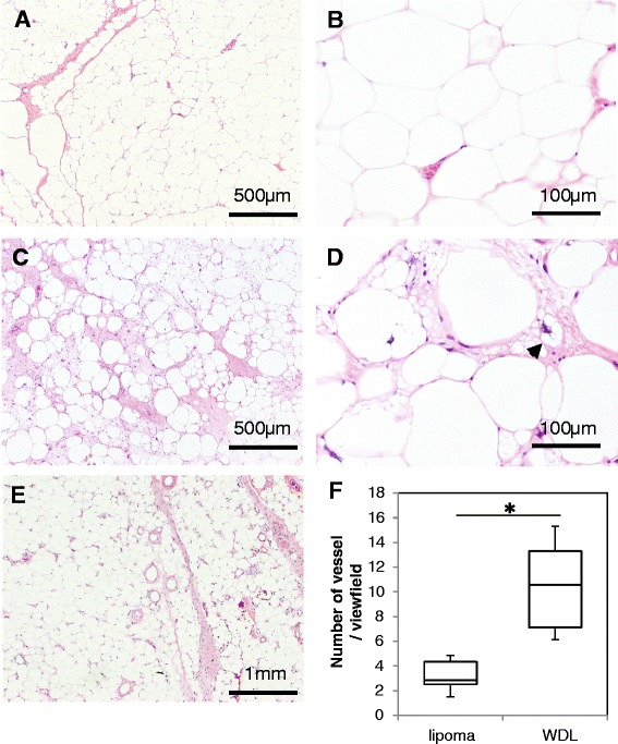
Forty-eight lipomas and 12 ALTs were included. The mean age, location and depth of the tumor as well as the compartment were not significantly different between the 2 groups. To evaluate the vascularity of the tumors, the average number of intratumoral vessels on pathological sections was calculated and compared between cases of lipoma and ALT.

RESULTS

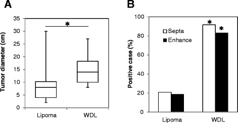
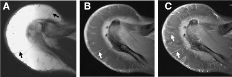
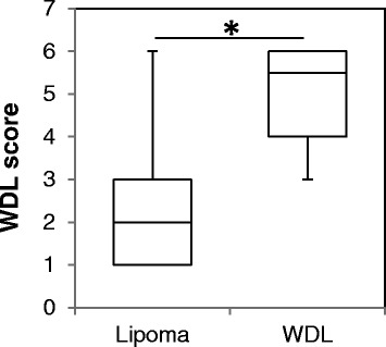
The tumor size was significantly larger in ALT cases than in lipoma cases (P < 0.001). Magnetic resonance imaging (MRI) revealed septal structures in 91.6% of ALTs, whereas 20.8% of lipomas showed septa. Contrast enhancement in MRI was found significantly more often in ALTs (81.2%) than in lipomas (18.8%) (P < 0.001). We created a "ALT score" to discriminate between lipoma and ALT (0-6 points). ALT cases gave significantly higher point values (average 5.1 points) than lipoma cases (average 1.7 points) (P < 0.001). We found a significantly increased number of vessels in cases of ALT than in cases of lipoma (P = 0.001).

CONCLUSIONS

Our ALT score may help surgeons to differentiate a suspected ALT from a lipoma and could recommend a marginal resection in cases of suspected ALT. Increased intratumoral vascularity in ALT is reflected in the MRI findings and may play a key role in the acquisition of a malignant phenotype in adipocytic tumors.

摘要

背景

高分化脂肪肉瘤(WDL)/非典型脂肪瘤性肿瘤(ALT)被认为是一种低级别恶性肿瘤,很少发生转移,但因其可能出现复发或去分化,故应密切随访。人们认识到WDL和ALT本质上是同义词,描述的是形态学和核型均相同的病变,且行为上的部位特异性差异仅与手术可切除性有关。由于脂肪瘤和ALT的临床及影像特征非常相似,术前对二者进行鉴别诊断已得到充分研究。我们评估了可能区分ALT与脂肪瘤的因素,并验证了一种用于诊断这两种肿瘤类型的初步评分系统。

方法

纳入48例脂肪瘤和12例ALT。两组在肿瘤的平均年龄、位置、深度以及分区方面无显著差异。为评估肿瘤的血管情况,计算并比较了脂肪瘤和ALT病例病理切片上瘤内血管的平均数量。

结果

ALT病例的肿瘤大小显著大于脂肪瘤病例(P < 0.001)。磁共振成像(MRI)显示91.6%的ALT有分隔结构,而20.8%的脂肪瘤有分隔。MRI上ALT的对比增强明显多于脂肪瘤(81.2% 对18.8%)(P < 0.001)。我们创建了一个“ALT评分”来区分脂肪瘤和ALT(0 - 6分)。ALT病例的得分显著高于脂肪瘤病例(平均分5.1分对1.7分)(P < 0.001)。我们发现ALT病例的血管数量显著多于脂肪瘤病例(P = 0.001)。

结论

我们的ALT评分可能有助于外科医生将疑似ALT与脂肪瘤区分开来,并在疑似ALT的病例中建议进行边缘切除。ALT中瘤内血管增多在MRI表现中有所体现,可能在脂肪细胞性肿瘤获得恶性表型中起关键作用。

https://cdn.ncbi.nlm.nih.gov/pmc/blobs/e752/4340111/0d67a319c6f0/12891_2015_491_Fig5_HTML.jpg
https://cdn.ncbi.nlm.nih.gov/pmc/blobs/e752/4340111/52106db5ba6a/12891_2015_491_Fig1_HTML.jpg
https://cdn.ncbi.nlm.nih.gov/pmc/blobs/e752/4340111/d9a158dd8010/12891_2015_491_Fig2_HTML.jpg
https://cdn.ncbi.nlm.nih.gov/pmc/blobs/e752/4340111/a08e8bcfdf40/12891_2015_491_Fig3_HTML.jpg
https://cdn.ncbi.nlm.nih.gov/pmc/blobs/e752/4340111/cc2aee7fefe0/12891_2015_491_Fig4_HTML.jpg
https://cdn.ncbi.nlm.nih.gov/pmc/blobs/e752/4340111/0d67a319c6f0/12891_2015_491_Fig5_HTML.jpg

相似文献

1
Differentiation of lipoma and atypical lipomatous tumor by a scoring system: implication of increased vascularity on pathogenesis of liposarcoma.通过评分系统鉴别脂肪瘤和非典型脂肪瘤性肿瘤:血管增多对脂肪肉瘤发病机制的影响
BMC Musculoskelet Disord. 2015 Feb 22;16:36. doi: 10.1186/s12891-015-0491-8.
2
MDM2 Amplification in Problematic Lipomatous Tumors: Analysis of FISH Testing Criteria.疑难脂肪性肿瘤中的MDM2基因扩增:荧光原位杂交检测标准分析
Am J Surg Pathol. 2015 Oct;39(10):1433-9. doi: 10.1097/PAS.0000000000000468.
3
Developing a clinical scoring system to differentiate deep-seated atypical lipomatous tumor from lipoma of soft tissue.制定一种临床评分系统以区分深部非典型性脂肪肉瘤与软组织脂肪瘤。
Asian J Surg. 2019 Aug;42(8):832-838. doi: 10.1016/j.asjsur.2018.12.012. Epub 2019 Jan 30.
4
Differentiating atypical lipomatous tumors from lipomas with magnetic resonance imaging: a comparison with MDM2 gene amplification status.用磁共振成像区分非典型性脂肪肿瘤与脂肪瘤:与 MDM2 基因扩增状态的比较。
BMC Cancer. 2019 Apr 3;19(1):309. doi: 10.1186/s12885-019-5524-5.
5
Equivalent cross-relaxation rate imaging positively correlates with pathological grade and cell density of adipocytic tumors.等效交叉弛豫率成像与脂肪细胞肿瘤的病理分级和细胞密度呈正相关。
Magn Reson Imaging. 2014 Apr;32(3):206-10. doi: 10.1016/j.mri.2013.12.004. Epub 2013 Dec 18.
6
MRI characteristics of lipoma and atypical lipomatous tumor/well-differentiated liposarcoma: retrospective comparison with histology and MDM2 gene amplification.MRI 特征在脂肪瘤和非典型性脂肪肉瘤/高分化脂肪肉瘤中的表现:与组织学和 MDM2 基因扩增的回顾性比较。
Skeletal Radiol. 2013 May;42(5):635-47. doi: 10.1007/s00256-012-1517-z. Epub 2012 Sep 18.
7
Differential diagnosis of benign peripheral lipoma from well-differentiated liposarcoma on MR imaging: is comparison of margins and internal characteristics useful?磁共振成像上良性周围脂肪瘤与高分化脂肪肉瘤的鉴别诊断:边缘及内部特征的比较有用吗?
AJR Am J Roentgenol. 2003 Jun;180(6):1689-94. doi: 10.2214/ajr.180.6.1801689.
8
Image-Guided Core Needle Biopsy of Adipocytic Tumors: Diagnostic Accuracy and Concordance With Final Surgical Pathology.影像引导下的脂肪细胞肿瘤核心针活检:诊断准确性及与最终外科病理的一致性。
AJR Am J Roentgenol. 2021 Apr;216(4):997-1002. doi: 10.2214/AJR.20.23080. Epub 2021 Feb 24.
9
Review of large deep lipomatous tumours.大型深部脂肪瘤性肿瘤综述。
ANZ J Surg. 2007 Jul;77(7):524-9. doi: 10.1111/j.1445-2197.2007.04042.x.
10
Contrast-enhanced ultrasound reveals perfusion differences between benign lipoma and semi-malignant atypical lipomatous tumors: A prospective clinical study.超声造影显示良性脂肪瘤与半恶性非典型性脂肪瘤肿瘤之间的灌注差异:一项前瞻性临床研究。
Ultraschall Med. 2024 Oct;45(5):509-518. doi: 10.1055/a-2189-5412. Epub 2023 Oct 11.

引用本文的文献

1
Atypical Lipomatous Tumours vs. Lipomas: A Multimodal Diagnostic Approach.非典型脂肪瘤性肿瘤与脂肪瘤:多模态诊断方法
Diagnostics (Basel). 2025 Jun 17;15(12):1538. doi: 10.3390/diagnostics15121538.
2
MRI of atypical lipomatous tumor: does contrast help? A multicenter study.非典型脂肪瘤性肿瘤的磁共振成像:造影剂有帮助吗?一项多中心研究。
Skeletal Radiol. 2025 Jun 5. doi: 10.1007/s00256-025-04957-8.
3
Dedifferentiated liposarcoma of the anterior mediastinum: An uncommon case presentation and complex diagnosis.前纵隔去分化脂肪肉瘤:1例罕见病例报告及复杂诊断

本文引用的文献

1
Deep-seated huge hibernoma of soft tissue: a rare differential diagnosis of atypical lipomatous tumor/well differentiated liposarcoma.深部软组织巨大冬眠瘤:非典型脂肪瘤性肿瘤/高分化脂肪肉瘤的一种罕见鉴别诊断。
Int J Clin Exp Pathol. 2013 Sep 15;6(10):2178-84. eCollection 2013.
2
Primary (de novo) dedifferentiated liposarcoma in the extremities: a multi-institution Tohoku Musculoskeletal Tumor Society study of 18 cases in northern Japan.四肢原发性(从头发生)去分化脂肪肉瘤:日本北部 18 例东北肌肉骨骼肿瘤学会多机构研究。
Jpn J Clin Oncol. 2011 Sep;41(9):1094-100. doi: 10.1093/jjco/hyr098. Epub 2011 Jul 26.
3
Well-differentiated and dedifferentiated liposarcomas.
Radiol Case Rep. 2025 Apr 3;20(6):3076-3080. doi: 10.1016/j.radcr.2025.03.035. eCollection 2025 Jun.
4
Management Algorithm for Atypical Lipomatous Tumours: A Retrospective Case Series.非典型脂肪瘤性肿瘤的管理算法:一项回顾性病例系列研究
Cureus. 2025 Jan 13;17(1):e77384. doi: 10.7759/cureus.77384. eCollection 2025 Jan.
5
The Lipid Enigma: A Case Report Highlighting Diagnostic Challenges in Adipocytic Tumors.脂质之谜:一例突出脂肪细胞肿瘤诊断挑战的病例报告
Cureus. 2024 Oct 10;16(10):e71233. doi: 10.7759/cureus.71233. eCollection 2024 Oct.
6
Mediastinal liposarcoma: Case report with radiology review.纵隔脂肪肉瘤:病例报告及影像学回顾
Radiol Case Rep. 2024 May 18;19(8):3294-3300. doi: 10.1016/j.radcr.2024.04.054. eCollection 2024 Aug.
7
UK guidelines for the management of soft tissue sarcomas.英国软组织肉瘤管理指南。
Br J Cancer. 2025 Jan;132(1):11-31. doi: 10.1038/s41416-024-02674-y. Epub 2024 May 11.
8
Differential Diagnosis of Lipomatous Tumors Using F-Fluorodeoxygulcose Positron Emission Tomography/Computed Tomography: A Retrospective Observational Study.使用F-氟脱氧葡萄糖正电子发射断层扫描/计算机断层扫描对脂肪瘤性肿瘤进行鉴别诊断:一项回顾性观察研究。
Cancer Diagn Progn. 2024 Mar 3;4(2):141-146. doi: 10.21873/cdp.10300. eCollection 2024 Mar-Apr.
9
The Predictive Role of the Histopathological Scoring System in Adipose Tumors-Lipoma, Atypical Lipomatous Tumor, and Liposarcoma.组织病理学评分系统在脂肪性肿瘤(脂肪瘤、非典型脂肪瘤性肿瘤和脂肪肉瘤)中的预测作用
Diagnostics (Basel). 2023 Dec 5;13(24):3606. doi: 10.3390/diagnostics13243606.
10
Diagnostic accuracy of CT and MR features for detecting atypical lipomatous tumors and malignant liposarcomas: a systematic review and meta-analysis.CT和MR特征对非典型脂肪瘤性肿瘤和恶性脂肪肉瘤的诊断准确性:一项系统评价和荟萃分析
Eur Radiol. 2023 Dec;33(12):8605-8616. doi: 10.1007/s00330-023-09916-2. Epub 2023 Jul 13.
高分化和去分化脂肪肉瘤。
Virchows Arch. 2010 Feb;456(2):167-79. doi: 10.1007/s00428-009-0815-x. Epub 2009 Aug 18.
4
False-positive diagnosis of metastasis on positron emission tomography-computed tomography imaging due to hibernoma.由于冬眠瘤导致正电子发射断层扫描-计算机断层扫描成像出现转移的假阳性诊断。
J Clin Oncol. 2009 Feb 20;27(6):994-5. doi: 10.1200/JCO.2008.19.4977. Epub 2008 Dec 29.
5
Tumor dormancy due to failure of angiogenesis: role of the microenvironment.血管生成失败导致的肿瘤休眠:微环境的作用
Clin Exp Metastasis. 2009;26(1):51-60. doi: 10.1007/s10585-008-9176-0. Epub 2008 Jun 18.
6
Magnetic resonance imaging of lipoma and atypical lipomatous tumour/well-differentiated liposarcoma: observer performance using T1-weighted and fluid-sensitive MRI.脂肪瘤和非典型脂肪瘤性肿瘤/高分化脂肪肉瘤的磁共振成像:使用T1加权和液体敏感型MRI的观察者表现
J Med Imaging Radiat Oncol. 2008 Feb;52(1):44-8. doi: 10.1111/j.1440-1673.2007.01910.x.
7
Conservative surgery for well-differentiated liposarcomas of the extremities adjacent to major neurovascular structures.对邻近主要神经血管结构的四肢高分化脂肪肉瘤进行保肢手术。
Surg Oncol. 2006 Nov;15(3):167-71. doi: 10.1016/j.suronc.2006.11.004. Epub 2006 Dec 20.
8
Clinical outcomes of deep atypical lipomas (well-differentiated lipoma-like liposarcomas) of the extremities.四肢深部非典型脂肪瘤(高分化脂肪瘤样脂肪肉瘤)的临床结局
ANZ J Surg. 2005 Sep;75(9):803-6. doi: 10.1111/j.1445-2197.2005.03519.x.
9
Deep-seated, well differentiated lipomatous tumors of the chest wall and extremities: the role of cytogenetics in classification and prognostication.胸壁和四肢深部、高分化脂肪性肿瘤:细胞遗传学在分类和预后评估中的作用
Cancer. 2005 Jan 15;103(2):409-16. doi: 10.1002/cncr.20779.
10
Lipomas, lipoma variants, and well-differentiated liposarcomas (atypical lipomas): results of MRI evaluations of 126 consecutive fatty masses.脂肪瘤、脂肪瘤变体及高分化脂肪肉瘤(非典型脂肪瘤):126例连续脂肪性肿块的MRI评估结果
AJR Am J Roentgenol. 2004 Mar;182(3):733-9. doi: 10.2214/ajr.182.3.1820733.