Changrob Siriruk, Leepiyasakulchai Chaniya, Tsuboi Takafumi, Cheng Yang, Lim Chae Seung, Chootong Patchanee, Han Eun-Taek
Department of Clinical Microbiology and Applied Technology, Faculty of Medical Technology, Mahidol University, Bangkok, Thailand.
Division of Malaria Research Proteo-Science Center, Ehime University, Matsuyama, Ehime, 790-8577, Japan.
Malar J. 2015 Apr 15;14:159. doi: 10.1186/s12936-015-0681-8.
Plasmodium vivax merozoite surface protein-1 paralog (PvMSP1P) is a glycosylphosphatidylinositol-anchored protein expressed on the merozoite surface. This molecule is a target of natural immunity, as high anti-MSP1P-19 antibody levels were detected during P. vivax infection and the antibody inhibited PvMSP1P-erythrocyte binding. Recombinant PvMSP1P antigen results in production of a significant Th1 cytokine response in immunized mice. The present study was performed to characterize natural cellular immunity against PvMSP1P-19 and PvDBP region II in acute and recovery P. vivax infection.
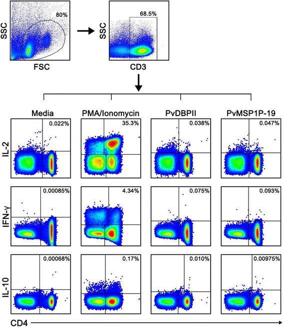
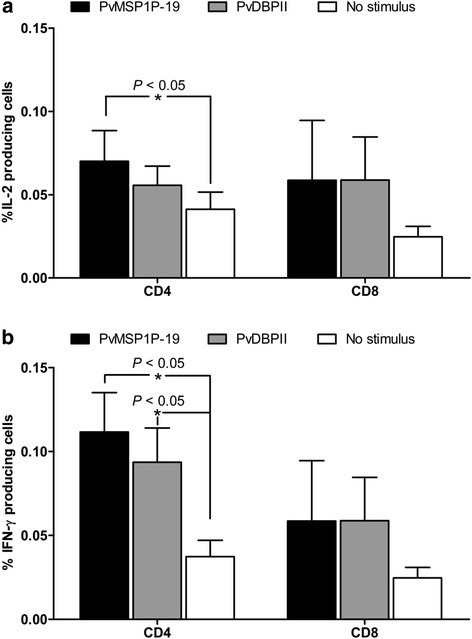
Peripheral blood mononuclear cells (PBMCs) from acute and recovery P. vivax infection were obtained for lymphocyte proliferation assay upon PvMSP1P-19 and PvDBP region II antigen stimulation. The culture supernatant was examined for the presence of the cytokines IL-2, TNF, IFN-γ and IL-10 by enzyme-linked immunosorbent assay (ELISA). To determine whether Th1 or Th2 have a memory response against PvMSP1P-19 and PvDBPII protein antigen, PBMCs from subjects who had recovered from P. vivax infection 8-10 weeks prior to the study were obtained for lymphocyte proliferation assay. Cytokine-producing cells were analysed by flow cytometry.
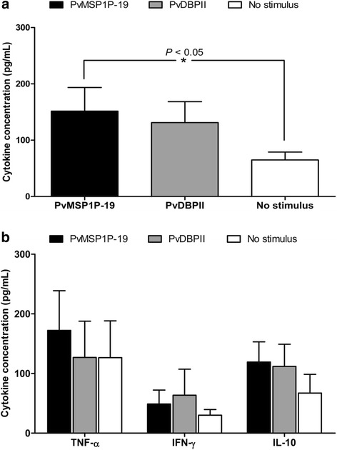
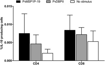
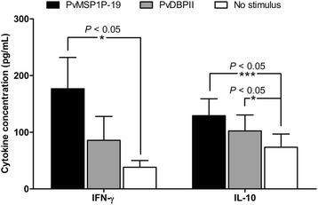
IL-2 was detected at high levels in lymphocyte cultures from acutely infected P. vivax patients upon PvMSP1P-19 stimulation. Analysis of the Th1 or Th2 memory response in PBMC cultures from subjects who had recovered from P. vivax infection showed significantly elevated levels of PvMSP1P-19 and PvDBPII-specific IFN-γ-producing cells (P < 0.05). Interestingly, the response of IFN-γ-producing cells in PvMSP1P stimulation was fourfold greater in recovered subjects than that in acute-infection patients. CD4(+) T cells were the major cell phenotype involved in the response to PvMSP1P-19 and PvDBPII antigen.
PvMSP1P-19 strongly induces a specific cellular immune response for protection against P. vivax compared with PvDBPII as the antigen induces activation of IFN-γ-producing effector cells following natural P. vivax exposure. Upon stimulation, PvMSP1P-19 has the potential to activate the recall response of Th1 effector memory cells that play a role in killing the parasite.
间日疟原虫裂殖子表面蛋白-1旁系同源物(PvMSP1P)是一种糖基磷脂酰肌醇锚定蛋白,表达于裂殖子表面。该分子是天然免疫的靶点,因为在间日疟原虫感染期间检测到高抗MSP1P-19抗体水平,且该抗体可抑制PvMSP1P与红细胞的结合。重组PvMSP1P抗原在免疫小鼠中可诱导显著的Th1细胞因子反应。本研究旨在表征急性和恢复期间日疟原虫感染中针对PvMSP1P-19和PvDBP区域II的天然细胞免疫。
获取急性和恢复期间日疟原虫感染患者的外周血单个核细胞(PBMC),在PvMSP1P-19和PvDBP区域II抗原刺激下进行淋巴细胞增殖试验。通过酶联免疫吸附测定(ELISA)检测培养上清液中细胞因子IL-2、TNF、IFN-γ和IL-10的存在情况。为确定Th1或Th2是否对PvMSP1P-19和PvDBPII蛋白抗原有记忆反应,获取在研究前8 - 10周从间日疟原虫感染中恢复的受试者的PBMC进行淋巴细胞增殖试验。通过流式细胞术分析产生细胞因子的细胞。
在PvMSP1P-19刺激下,急性感染间日疟原虫患者的淋巴细胞培养物中检测到高水平的IL-2。对从间日疟原虫感染中恢复的受试者的PBMC培养物中Th1或Th2记忆反应的分析显示,PvMSP1P-19和PvDBPII特异性产生IFN-γ的细胞水平显著升高(P < 0.05)。有趣的是,在恢复的受试者中,PvMSP1P刺激下产生IFN-γ的细胞反应比急性感染患者高四倍。CD4(+) T细胞是参与对PvMSP1P-19和PvDBPII抗原反应的主要细胞表型。
与PvDBPII相比,PvMSP1P-19强烈诱导针对间日疟原虫的特异性细胞免疫反应,因为该抗原在间日疟原虫自然暴露后可诱导产生IFN-γ的效应细胞活化。受到刺激后,PvMSP1P-19有可能激活Th1效应记忆细胞的回忆反应,这些细胞在杀死寄生虫中发挥作用。