• 文献检索
  • 文档翻译
  • 深度研究
  • 学术资讯
  • Suppr Zotero 插件Zotero 插件
  • 邀请有礼
  • 套餐&价格
  • 历史记录
应用&插件
Suppr Zotero 插件Zotero 插件浏览器插件Mac 客户端Windows 客户端微信小程序
定价
高级版会员购买积分包购买API积分包
服务
文献检索文档翻译深度研究API 文档MCP 服务
关于我们
关于 Suppr公司介绍联系我们用户协议隐私条款
关注我们

Suppr 超能文献

核心技术专利:CN118964589B侵权必究
粤ICP备2023148730 号-1Suppr @ 2026

文献检索

告别复杂PubMed语法,用中文像聊天一样搜索,搜遍4000万医学文献。AI智能推荐,让科研检索更轻松。

立即免费搜索

文件翻译

保留排版,准确专业,支持PDF/Word/PPT等文件格式,支持 12+语言互译。

免费翻译文档

深度研究

AI帮你快速写综述,25分钟生成高质量综述,智能提取关键信息,辅助科研写作。

立即免费体验

人类神经干细胞磁标记及偏侧帕金森病大鼠脑MRI可视化的优化

Optimization of the magnetic labeling of human neural stem cells and MRI visualization in the hemiparkinsonian rat brain.

作者信息

Ramos-Gómez Milagros, Seiz Emma G, Martínez-Serrano Alberto

机构信息

Centre for Biomedical Technology, Polytechnic University of Madrid, 28223, Madrid, Spain.

Biomedical Research Networking Center in Bioengineering Biomaterials and Nanomedicine (CIBER-BBN), Madrid, Spain.

出版信息

J Nanobiotechnology. 2015 Mar 5;13:20. doi: 10.1186/s12951-015-0078-4.

DOI:10.1186/s12951-015-0078-4
PMID:25890124
原文链接:https://pmc.ncbi.nlm.nih.gov/articles/PMC4416262/
Abstract

BACKGROUND

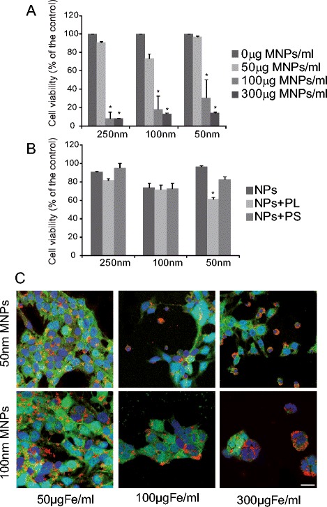
Magnetic resonance imaging is the ideal modality for non-invasive in vivo cell tracking allowing for longitudinal studies over time. Cells labeled with superparamagnetic iron oxide nanoparticles have been shown to induce sufficient contrast for in vivo magnetic resonance imaging enabling the in vivo analysis of the final location of the transplanted cells. For magnetic nanoparticles to be useful, a high internalization efficiency of the particles is required without compromising cell function, as well as validation of the magnetic nanoparticles behaviour inside the cells.

RESULTS

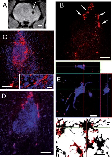
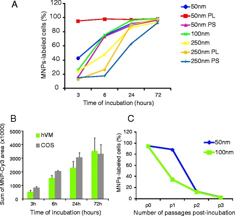
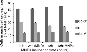
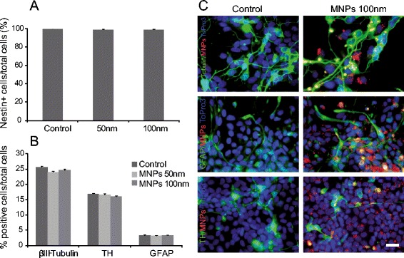
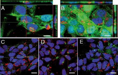
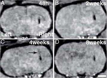
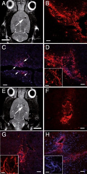
In this work, we report the development, optimization and validation of an efficient procedure to label human neural stem cells with commercial nanoparticles in the absence of transfection agents. Magnetic nanoparticles used here do not affect cell viability, cell morphology, cell differentiation or cell cycle dynamics. Moreover, human neural stem cells progeny labeled with magnetic nanoparticles are easily and non-invasively detected long time after transplantation in a rat model of Parkinson's disease (up to 5 months post-grafting) by magnetic resonance imaging.

CONCLUSIONS

These findings support the use of commercial MNPs to track cells for short- and mid-term periods after transplantation for studies of brain cell replacement therapy. Nevertheless, long-term MR images should be interpreted with caution due to the possibility that some MNPs may be expelled from the transplanted cells and internalized by host microglial cells.

摘要

背景

磁共振成像(MRI)是用于体内细胞无创追踪的理想方式,可进行长期的纵向研究。已证实,用超顺磁性氧化铁纳米颗粒标记的细胞能在体内磁共振成像中产生足够的对比度,从而实现对移植细胞最终位置的体内分析。要使磁性纳米颗粒发挥作用,需要其具有较高的内化效率且不影响细胞功能,同时还需验证磁性纳米颗粒在细胞内的行为。

结果

在本研究中,我们报告了一种在无转染剂情况下用商业纳米颗粒标记人神经干细胞的高效方法的开发、优化及验证。此处使用的磁性纳米颗粒不影响细胞活力、细胞形态、细胞分化或细胞周期动力学。此外,在帕金森病大鼠模型中移植后很长时间(移植后长达5个月),通过磁共振成像可轻松且无创地检测到用磁性纳米颗粒标记的人神经干细胞后代。

结论

这些发现支持使用商业磁性纳米颗粒在移植后的短期和中期追踪细胞,用于脑细胞替代治疗研究。然而,由于一些磁性纳米颗粒可能从移植细胞中排出并被宿主小胶质细胞内化,因此对长期磁共振图像的解读应谨慎。

https://cdn.ncbi.nlm.nih.gov/pmc/blobs/3d92/4416262/584ee02a95d6/12951_2015_78_Fig8_HTML.jpg
https://cdn.ncbi.nlm.nih.gov/pmc/blobs/3d92/4416262/f6463a60ffe0/12951_2015_78_Fig1_HTML.jpg
https://cdn.ncbi.nlm.nih.gov/pmc/blobs/3d92/4416262/535906ae7939/12951_2015_78_Fig2_HTML.jpg
https://cdn.ncbi.nlm.nih.gov/pmc/blobs/3d92/4416262/42104c6dec07/12951_2015_78_Fig3_HTML.jpg
https://cdn.ncbi.nlm.nih.gov/pmc/blobs/3d92/4416262/c4ad8a942378/12951_2015_78_Fig4_HTML.jpg
https://cdn.ncbi.nlm.nih.gov/pmc/blobs/3d92/4416262/a753f517f919/12951_2015_78_Fig5_HTML.jpg
https://cdn.ncbi.nlm.nih.gov/pmc/blobs/3d92/4416262/17b4c31a8aa6/12951_2015_78_Fig6_HTML.jpg
https://cdn.ncbi.nlm.nih.gov/pmc/blobs/3d92/4416262/88708df56e55/12951_2015_78_Fig7_HTML.jpg
https://cdn.ncbi.nlm.nih.gov/pmc/blobs/3d92/4416262/584ee02a95d6/12951_2015_78_Fig8_HTML.jpg

相似文献

1
Optimization of the magnetic labeling of human neural stem cells and MRI visualization in the hemiparkinsonian rat brain.人类神经干细胞磁标记及偏侧帕金森病大鼠脑MRI可视化的优化
J Nanobiotechnology. 2015 Mar 5;13:20. doi: 10.1186/s12951-015-0078-4.
2
A DNA hybridization system for labeling of neural stem cells with SPIO nanoparticles for MRI monitoring post-transplantation.一种用于用超顺磁氧化铁纳米颗粒标记神经干细胞的 DNA 杂交系统,用于移植后 MRI 监测。
Biomaterials. 2015 Jun;54:158-67. doi: 10.1016/j.biomaterials.2015.03.017. Epub 2015 Apr 3.
3
In Vivo Tracking of Human Neural Progenitor Cells in the Rat Brain Using Magnetic Resonance Imaging Is Not Enhanced by Ferritin Expression.使用磁共振成像对大鼠脑中人类神经祖细胞进行体内追踪时,铁蛋白表达并未增强追踪效果。
Cell Transplant. 2016;25(3):575-92. doi: 10.3727/096368915X688614. Epub 2015 Jul 8.
4
D-mannose-Coating of Maghemite Nanoparticles Improved Labeling of Neural Stem Cells and Allowed Their Visualization by MRI after Transplantation in the Mouse Brain.磁赤铁矿纳米粒子的 D-甘露糖涂层改善了神经干细胞的标记效果,使其在移植到小鼠大脑后可通过 MRI 进行可视化。
Cell Transplant. 2019 May;28(5):553-567. doi: 10.1177/0963689719834304. Epub 2019 Jul 11.
5
Effects of the iron oxide nanoparticle Molday ION Rhodamine B on the viability and regenerative function of neural stem cells: relevance to clinical translation.氧化铁纳米颗粒Molday ION罗丹明B对神经干细胞活力和再生功能的影响:与临床转化的相关性。
Int J Nanomedicine. 2016 Apr 27;11:1731-48. doi: 10.2147/IJN.S102006. eCollection 2016.
6
Ex vivo magnetic resonance imaging of transplanted hepatocytes in a rat model of acute liver failure.在急性肝衰竭大鼠模型中移植肝细胞的离体磁共振成像。
Cell Transplant. 2014 Mar;23(3):329-43. doi: 10.3727/096368913X663596. Epub 2013 Feb 5.
7
Enhanced cellular uptake and long-term retention of chitosan-modified iron-oxide nanoparticles for MRI-based cell tracking.壳聚糖修饰的氧化铁纳米颗粒增强细胞摄取和长期保留用于基于 MRI 的细胞示踪。
Int J Nanomedicine. 2012;7:4613-23. doi: 10.2147/IJN.S28294. Epub 2012 Aug 22.
8
Superparamagnetic iron oxide nanoparticles as a tool to track mouse neural stem cells in vivo.超顺磁性氧化铁纳米颗粒作为在体内追踪小鼠神经干细胞的工具。
Mol Biol Rep. 2019 Feb;46(1):191-198. doi: 10.1007/s11033-018-4460-9. Epub 2018 Nov 12.
9
Mesenchymal stromal cell labeling by new uncoated superparamagnetic maghemite nanoparticles in comparison with commercial Resovist--an initial in vitro study.新型未包被超顺磁性磁赤铁矿纳米颗粒与市售Resovist对比标记间充质基质细胞——一项初步体外研究
Int J Nanomedicine. 2014 Nov 20;9:5355-72. doi: 10.2147/IJN.S66986. eCollection 2014.
10
Detection of viability of transplanted beta cells labeled with a novel contrast agent - polyvinylpyrrolidone-coated superparamagnetic iron oxide nanoparticles by magnetic resonance imaging.新型对比剂——聚乙烯吡咯烷酮包裹超顺磁性氧化铁纳米颗粒标记的移植胰岛细胞活力的磁共振成像检测。
Contrast Media Mol Imaging. 2012 Jan-Feb;7(1):35-44. doi: 10.1002/cmmi.461.

引用本文的文献

1
Cytotoxic effect and apoptotic induction of tricyclohexyltin p-methoxycinnamate on HT-29 colorectal cancer cells: Implications for anticancer therapeutics.对甲氧基肉桂酸三环己基锡对HT-29结肠癌细胞的细胞毒性作用及凋亡诱导:对抗癌治疗的意义。
J Adv Pharm Technol Res. 2025 Jan-Mar;16(1):1-5. doi: 10.4103/JAPTR.JAPTR_427_23. Epub 2025 Feb 14.
2
Tracking Neural Stem Cells in vivo: Achievements and Limitations.在体追踪神经干细胞:成就与局限。
Stem Cell Rev Rep. 2022 Jun;18(5):1774-1788. doi: 10.1007/s12015-022-10333-z. Epub 2022 Feb 5.
3
Importance of Nanoparticles for the Delivery of Antiparkinsonian Drugs.

本文引用的文献

1
Human ESC-derived dopamine neurons show similar preclinical efficacy and potency to fetal neurons when grafted in a rat model of Parkinson's disease.当移植到帕金森病大鼠模型中时,人胚胎干细胞衍生的多巴胺神经元显示出与胎儿神经元相似的临床前疗效和效力。
Cell Stem Cell. 2014 Nov 6;15(5):653-65. doi: 10.1016/j.stem.2014.09.017.
2
Influence of oxygen tension on dopaminergic differentiation of human fetal stem cells of midbrain and forebrain origin.氧张力对中脑和前脑来源的人胎儿干细胞多巴胺能分化的影响。
PLoS One. 2014 May 2;9(5):e96465. doi: 10.1371/journal.pone.0096465. eCollection 2014.
3
Human conditionally immortalized neural stem cells improve locomotor function after spinal cord injury in the rat.
纳米颗粒在抗帕金森病药物递送中的重要性。
Pharmaceutics. 2021 Apr 8;13(4):508. doi: 10.3390/pharmaceutics13040508.
4
Current Status of Stem Cell-Derived Therapies for Parkinson's Disease: From Cell Assessment and Imaging Modalities to Clinical Trials.帕金森病干细胞衍生疗法的现状:从细胞评估与成像方式到临床试验
Front Neurosci. 2020 Oct 16;14:558532. doi: 10.3389/fnins.2020.558532. eCollection 2020.
5
Cellular and molecular imaging for stem cell tracking in neurological diseases.用于神经疾病中干细胞示踪的细胞和分子成像。
Stroke Vasc Neurol. 2021 Mar;6(1):121-127. doi: 10.1136/svn-2020-000408. Epub 2020 Oct 29.
6
Magnetic resonance imaging of human neural stem cells in rodent and primate brain.磁共振成像在啮齿动物和灵长类动物脑内人类神经干细胞的应用。
Stem Cells Transl Med. 2021 Jan;10(1):83-97. doi: 10.1002/sctm.20-0126. Epub 2020 Aug 25.
7
Application of Iron Oxide Nanoparticles in the Diagnosis and Treatment of Neurodegenerative Diseases With Emphasis on Alzheimer's Disease.氧化铁纳米颗粒在神经退行性疾病诊断与治疗中的应用——以阿尔茨海默病为重点
Front Cell Neurosci. 2020 Feb 28;14:21. doi: 10.3389/fncel.2020.00021. eCollection 2020.
8
Comparative strategies for stem cell biodistribution in a preclinical study.在临床前研究中比较干细胞生物分布的策略。
Acta Pharmacol Sin. 2020 Apr;41(4):572-580. doi: 10.1038/s41401-019-0313-x. Epub 2019 Nov 8.
9
Imaging Gliomas with Nanoparticle-Labeled Stem Cells.用纳米颗粒标记的干细胞进行脑胶质瘤成像。
Chin Med J (Engl). 2018 Mar 20;131(6):721-730. doi: 10.4103/0366-6999.226900.
10
Stem Cell Tracking Technologies for Neurological Regenerative Medicine Purposes.用于神经再生医学目的的干细胞追踪技术
Stem Cells Int. 2017;2017:2934149. doi: 10.1155/2017/2934149. Epub 2017 Sep 12.
人类条件永生化神经干细胞可改善大鼠脊髓损伤后的运动功能。
Stem Cell Res Ther. 2013 Jun 7;4(3):68. doi: 10.1186/scrt219.
4
Functional maturation of hPSC-derived forebrain interneurons requires an extended timeline and mimics human neural development.人胚胎干细胞源性前脑内神经元的功能成熟需要延长时间,并模拟人类神经发育。
Cell Stem Cell. 2013 May 2;12(5):573-86. doi: 10.1016/j.stem.2013.04.005.
5
Gold- and silver nanoparticles affect the growth characteristics of human embryonic neural precursor cells.金纳米粒子和银纳米粒子影响人胚胎神经前体细胞的生长特性。
PLoS One. 2013;8(3):e58211. doi: 10.1371/journal.pone.0058211. Epub 2013 Mar 11.
6
Clonal human fetal ventral mesencephalic dopaminergic neuron precursors for cell therapy research.用于细胞治疗研究的克隆人胎儿腹侧中脑多巴胺能神经元前体细胞。
PLoS One. 2012;7(12):e52714. doi: 10.1371/journal.pone.0052714. Epub 2012 Dec 31.
7
Human midbrain precursors activate the expected developmental genetic program and differentiate long-term to functional A9 dopamine neurons in vitro. Enhancement by Bcl-X(L).人脑中脑前体细胞在体外激活预期的发育基因程序,并长期分化为功能性 A9 多巴胺神经元。Bcl-X(L) 可增强这种分化。
Exp Cell Res. 2012 Nov 15;318(19):2446-59. doi: 10.1016/j.yexcr.2012.07.018. Epub 2012 Aug 2.
8
Labeling stem cells with superparamagnetic iron oxide nanoparticles: analysis of the labeling efficacy by microscopy and magnetic resonance imaging.用超顺磁性氧化铁纳米颗粒标记干细胞:通过显微镜和磁共振成像分析标记效果
Methods Mol Biol. 2012;906:239-52. doi: 10.1007/978-1-61779-953-2_18.
9
Long term behavioral effects of functional dopaminergic neurons generated from human neural stem cells in the rat 6-OH-DA Parkinson's disease model. Effects of the forced expression of BCL-X(L).人神经干细胞诱导分化的功能性多巴胺能神经元在大鼠 6-OH-DA 帕金森病模型中长期的行为学效应。BCL-X(L)强制表达的影响。
Behav Brain Res. 2012 Jun 15;232(1):225-32. doi: 10.1016/j.bbr.2012.04.020. Epub 2012 Apr 17.
10
Self-assembling nanocomplexes by combining ferumoxytol, heparin and protamine for cell tracking by magnetic resonance imaging.通过将 ferumoxytol、肝素和鱼精蛋白组合成自组装纳米复合物,用于磁共振成像的细胞跟踪。
Nat Med. 2012 Feb 26;18(3):463-7. doi: 10.1038/nm.2666.