• 文献检索
  • 文档翻译
  • 深度研究
  • 学术资讯
  • Suppr Zotero 插件Zotero 插件
  • 邀请有礼
  • 套餐&价格
  • 历史记录
应用&插件
Suppr Zotero 插件Zotero 插件浏览器插件Mac 客户端Windows 客户端微信小程序
定价
高级版会员购买积分包购买API积分包
服务
文献检索文档翻译深度研究API 文档MCP 服务
关于我们
关于 Suppr公司介绍联系我们用户协议隐私条款
关注我们

Suppr 超能文献

核心技术专利:CN118964589B侵权必究
粤ICP备2023148730 号-1Suppr @ 2026

文献检索

告别复杂PubMed语法,用中文像聊天一样搜索,搜遍4000万医学文献。AI智能推荐,让科研检索更轻松。

立即免费搜索

文件翻译

保留排版,准确专业,支持PDF/Word/PPT等文件格式,支持 12+语言互译。

免费翻译文档

深度研究

AI帮你快速写综述,25分钟生成高质量综述,智能提取关键信息,辅助科研写作。

立即免费体验

肿瘤坏死因子诱导基因6促进急性肝损伤小鼠的肝脏再生。

Tumor necrosis factor-inducible gene 6 promotes liver regeneration in mice with acute liver injury.

作者信息

Wang Sihyung, Lee Ji-Seon, Hyun Jeongeun, Kim Jieun, Kim Seung U, Cha Hyuk-Jin, Jung Youngmi

机构信息

Department of Intergrated Biological Science, Pusan National University, 63-2 Pusandaehak-ro, Kumjeong-gu, Pusan, 609-735, Korea.

Department of Life Science, Sogang University, Seoul, 121-742, Korea.

出版信息

Stem Cell Res Ther. 2015 Mar 11;6(1):20. doi: 10.1186/s13287-015-0019-z.

DOI:10.1186/s13287-015-0019-z
PMID:25890163
原文链接:https://pmc.ncbi.nlm.nih.gov/articles/PMC4396561/
Abstract

INTRODUCTION

Tumor necrosis factor-inducible gene 6 protein (TSG-6), one of the cytokines released by human mesenchymal stem/stromal cells (hMSC), has an anti-inflammatory effect and alleviates several pathological conditions; however, the hepatoprotective potential of TSG-6 remains unclear. We investigated whether TSG-6 promoted liver regeneration in acute liver failure.

METHODS

The immortalized hMSC (B10) constitutively over-expressing TSG-6 or empty plasmid (NC: Negative Control) were established, and either TSG-6 or NC-conditioned medium (CM) was intraperitoneally injected into mice with acute liver damage caused by CCl4. Mice were sacrificed at 3 days post-CM treatment.

RESULTS

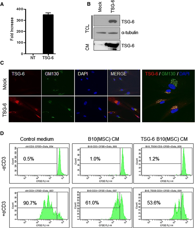
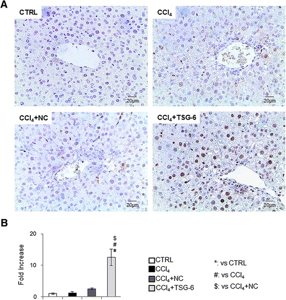
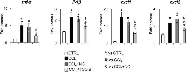
Higher expression and the immunosuppressive activity of TSG-6 were observed in CM from TSG-6-hMSC. The obvious histomorphological liver injury and increased level of liver enzymes were shown in CCl4-treated mice with or without NC-CM, whereas those observations were markedly ameliorated in TSG-6-CM-treated mice with CCl4. Ki67-positive hepatocytic cells were accumulated in the liver of the CCl4+TSG-6 group. RNA analysis showed the decrease in both of inflammation markers, tnfα, il-1β, cxcl1 and cxcl2, and fibrotic markers, tgf-β1, α-sma and collagen α1, in the CCl4+TSG-6 group, compared to the CCl4 or the CCl4+NC group. Protein analysis confirmed the lower expression of TGF-β1 and α-SMA in the CCl4+TSG-6 than the CCl4 or the CCl4+NC group. Immunostaining for α-SMA also revealed the accumulation of the activated hepatic stellate cells in the livers of mice in the CCl4 and CCl4+NC groups, but not in the livers of mice from the CCl4+TSG-6 group. The cultured LX2 cells, human hepatic stellate cell line, in TSG-6-CM showed the reduced expression of fibrotic markers, tgf-β1, vimentin and collagen α1, whereas the addition of the TSG-6 antibody neutralized the inhibitory effect of TSG-6 on the activation of LX2 cells. In addition, cytoplasmic lipid drops, the marker of inactivated hepatic stellate cell, were detected in TSG-6-CM-cultured LX2 cells, only. The suppressed TSG-6 activity by TSG-6 antibody attenuated the restoration process in livers of TSG-6-CM-treated mice with CCl4.

CONCLUSIONS

These results demonstrated that TSG-6 contributed to the liver regeneration by suppressing the activation of hepatic stellate cells in CCl4-treated mice, suggesting the therapeutic potential of TSG-6 for acute liver failure.

摘要

引言

肿瘤坏死因子诱导基因6蛋白(TSG - 6)是人间充质干/基质细胞(hMSC)释放的细胞因子之一,具有抗炎作用并能缓解多种病理状况;然而,TSG - 6的肝保护潜力仍不清楚。我们研究了TSG - 6是否能促进急性肝衰竭中的肝脏再生。

方法

构建了组成性过表达TSG - 6或空质粒(NC:阴性对照)的永生化hMSC(B10),并将TSG - 6或NC条件培养基(CM)腹腔注射到由四氯化碳引起急性肝损伤的小鼠体内。在CM处理后3天处死小鼠。

结果

在来自TSG - 6 - hMSC的CM中观察到TSG - 6的更高表达和免疫抑制活性。在接受或未接受NC - CM的四氯化碳处理小鼠中均出现明显的组织形态学肝损伤和肝酶水平升高,而在接受四氯化碳处理并注射TSG - 6 - CM的小鼠中这些观察结果明显改善。Ki67阳性肝细胞在四氯化碳 + TSG - 6组的肝脏中积聚。RNA分析显示,与四氯化碳组或四氯化碳 + NC组相比,四氯化碳 + TSG - 6组中炎症标志物(肿瘤坏死因子α、白细胞介素 - 1β、趋化因子CXCL1和CXCL2)以及纤维化标志物(转化生长因子 - β1、α - 平滑肌肌动蛋白和胶原蛋白α1)均减少。蛋白质分析证实,四氯化碳 + TSG - 6组中转化生长因子 - β1和α - SMA的表达低于四氯化碳组或四氯化碳 + NC组。α - SMA免疫染色还显示,四氯化碳组和四氯化碳 + NC组小鼠肝脏中活化的肝星状细胞积聚,而四氯化碳 + TSG - 6组小鼠肝脏中未出现。在TSG - 6 - CM中培养的LX2细胞(人肝星状细胞系)显示纤维化标志物(转化生长因子 - β1、波形蛋白和胶原蛋白α1)表达降低,而添加TSG - 6抗体可中和TSG - 6对LX2细胞活化的抑制作用。此外,仅在TSG - 6 - CM培养的LX2细胞中检测到细胞质脂滴(失活肝星状细胞的标志物)。TSG - 6抗体抑制TSG - 6活性减弱了接受四氯化碳处理并注射TSG - 6 - CM的小鼠肝脏的恢复过程。

结论

这些结果表明,TSG - 6通过抑制四氯化碳处理小鼠肝星状细胞的活化促进肝脏再生,提示TSG - 6对急性肝衰竭具有治疗潜力。

https://cdn.ncbi.nlm.nih.gov/pmc/blobs/33f0/4396561/d86934ec99ab/13287_2015_19_Fig9_HTML.jpg
https://cdn.ncbi.nlm.nih.gov/pmc/blobs/33f0/4396561/d7091424c391/13287_2015_19_Fig1_HTML.jpg
https://cdn.ncbi.nlm.nih.gov/pmc/blobs/33f0/4396561/a54ba32ae1d2/13287_2015_19_Fig2_HTML.jpg
https://cdn.ncbi.nlm.nih.gov/pmc/blobs/33f0/4396561/1d98db4e3841/13287_2015_19_Fig3_HTML.jpg
https://cdn.ncbi.nlm.nih.gov/pmc/blobs/33f0/4396561/3b79be8adb08/13287_2015_19_Fig4_HTML.jpg
https://cdn.ncbi.nlm.nih.gov/pmc/blobs/33f0/4396561/6345b085ba04/13287_2015_19_Fig5_HTML.jpg
https://cdn.ncbi.nlm.nih.gov/pmc/blobs/33f0/4396561/ed0e39e6686c/13287_2015_19_Fig6_HTML.jpg
https://cdn.ncbi.nlm.nih.gov/pmc/blobs/33f0/4396561/3ca58712d320/13287_2015_19_Fig7_HTML.jpg
https://cdn.ncbi.nlm.nih.gov/pmc/blobs/33f0/4396561/637efc7528da/13287_2015_19_Fig8_HTML.jpg
https://cdn.ncbi.nlm.nih.gov/pmc/blobs/33f0/4396561/d86934ec99ab/13287_2015_19_Fig9_HTML.jpg

相似文献

1
Tumor necrosis factor-inducible gene 6 promotes liver regeneration in mice with acute liver injury.肿瘤坏死因子诱导基因6促进急性肝损伤小鼠的肝脏再生。
Stem Cell Res Ther. 2015 Mar 11;6(1):20. doi: 10.1186/s13287-015-0019-z.
2
Mesenchymal stem cells and their secreted molecules predominantly ameliorate fulminant hepatic failure and chronic liver fibrosis in mice respectively.间充质干细胞及其分泌分子分别主要改善小鼠暴发性肝衰竭和慢性肝纤维化。
J Transl Med. 2016 Feb 9;14:45. doi: 10.1186/s12967-016-0792-1.
3
Tumor necrosis factor-inducible gene 6 reprograms hepatic stellate cells into stem-like cells, which ameliorates liver damage in mouse.肿瘤坏死因子诱导基因 6 可将肝星状细胞重编程为干细胞样细胞,从而改善小鼠的肝损伤。
Biomaterials. 2019 Oct;219:119375. doi: 10.1016/j.biomaterials.2019.119375. Epub 2019 Jul 22.
4
The therapeutic potential of human umbilical mesenchymal stem cells from Wharton's jelly in the treatment of rat liver fibrosis.来自华通氏胶的人脐带间充质干细胞在大鼠肝纤维化治疗中的治疗潜力。
Liver Transpl. 2009 May;15(5):484-95. doi: 10.1002/lt.21715.
5
Dihydromyricetin alleviates carbon tetrachloride-induced acute liver injury via JNK-dependent mechanism in mice.二氢杨梅素通过JNK依赖性机制减轻小鼠四氯化碳诱导的急性肝损伤。
World J Gastroenterol. 2015 May 14;21(18):5473-81. doi: 10.3748/wjg.v21.i18.5473.
6
Cell-specific PPARγ deficiency establishes anti-inflammatory and anti-fibrogenic properties for this nuclear receptor in non-parenchymal liver cells.细胞特异性过氧化物酶体增殖物激活受体 γ 缺失赋予非实质细胞中该核受体抗炎和抗纤维化特性。
J Hepatol. 2013 Nov;59(5):1045-53. doi: 10.1016/j.jhep.2013.06.023. Epub 2013 Jul 2.
7
Cav-1 deficiency promotes liver fibrosis in carbon tetrachloride (CCl)-induced mice by regulation of oxidative stress and inflammation responses.Cav-1 缺失通过调节氧化应激和炎症反应促进四氯化碳 (CCl) 诱导的小鼠肝纤维化。
Biomed Pharmacother. 2018 Jun;102:26-33. doi: 10.1016/j.biopha.2018.03.016. Epub 2018 Mar 14.
8
Matricellular protein periostin contributes to hepatic inflammation and fibrosis.基质细胞衍生蛋白 periostin 有助于肝脏炎症和纤维化。
Am J Pathol. 2015 Mar;185(3):786-97. doi: 10.1016/j.ajpath.2014.11.002. Epub 2014 Dec 23.
9
Phycocyanobilin accelerates liver regeneration and reduces mortality rate in carbon tetrachloride-induced liver injury mice.藻蓝胆素可加速四氯化碳诱导的肝损伤小鼠的肝脏再生并降低死亡率。
World J Gastroenterol. 2015 May 14;21(18):5465-72. doi: 10.3748/wjg.v21.i18.5465.
10
C-C motif chemokine receptor 9 positive macrophages activate hepatic stellate cells and promote liver fibrosis in mice.C-C 基序趋化因子受体 9 阳性巨噬细胞激活肝星状细胞并促进小鼠肝纤维化。
Hepatology. 2013 Jul;58(1):337-50. doi: 10.1002/hep.26351. Epub 2013 May 31.

引用本文的文献

1
CRISPR-dCas9 Activation of TSG-6 in MSCs Modulates the Cargo of MSC-Derived Extracellular Vesicles and Attenuates Inflammatory Responses in Human Intervertebral Disc Cells In Vitro.CRISPR-dCas9激活间充质干细胞中的TSG-6可调节间充质干细胞衍生的细胞外囊泡的成分,并在体外减轻人椎间盘细胞中的炎症反应。
Cell Mol Bioeng. 2025 Feb 5;18(1):83-98. doi: 10.1007/s12195-025-00843-4. eCollection 2025 Feb.
2
TFAIP6 facilitates hepatocellular carcinoma cell glycolysis through upregulating c-myc/PKM2 axis.TFAIP6通过上调c-myc/PKM2轴促进肝癌细胞糖酵解。
Heliyon. 2024 May 11;10(10):e30959. doi: 10.1016/j.heliyon.2024.e30959. eCollection 2024 May 30.
3

本文引用的文献

1
Hedgehog signaling influences gender-specific response of liver to radiation in mice.刺猬信号通路影响小鼠肝脏对辐射的性别特异性反应。
Hepatol Int. 2013 Oct;7(4):1065-74. doi: 10.1007/s12072-013-9461-0. Epub 2013 Jul 27.
2
Novel strategies for liver therapy using stem cells.使用干细胞进行肝脏治疗的新策略。
Gut. 2015 Jan;64(1):1-4. doi: 10.1136/gutjnl-2014-307480. Epub 2014 Sep 2.
3
Concise review: Therapeutic potential of mesenchymal stem cells for the treatment of acute liver failure and cirrhosis.简明综述:间充质干细胞治疗急性肝衰竭和肝硬化的治疗潜力。
TGF-β1 induces formation of TSG-6-enriched extracellular vesicles in fibroblasts which can prevent myofibroblast transformation by modulating Erk1/2 phosphorylation.
TGF-β1 诱导成纤维细胞形成富含 TSG-6 的细胞外囊泡,通过调节 Erk1/2 磷酸化来阻止肌成纤维细胞转化。
Sci Rep. 2024 May 29;14(1):12389. doi: 10.1038/s41598-024-62123-x.
4
Tumor necrosis factor-inducible gene 6 protein and its derived peptide ameliorate liver fibrosis by repressing CD44 activation in mice with alcohol-related liver disease.肿瘤坏死因子诱导基因 6 蛋白及其衍生肽通过抑制酒精性肝病小鼠 CD44 活化改善肝纤维化。
J Biomed Sci. 2024 May 24;31(1):54. doi: 10.1186/s12929-024-01042-5.
5
Improved therapeutic consistency and efficacy of CD317 MSCs through stabilizing TSG6 by PTX3.通过 PTX3 稳定 TSG6 可提高 CD317MSCs 的治疗一致性和疗效。
Stem Cell Res Ther. 2024 Mar 27;15(1):92. doi: 10.1186/s13287-024-03706-3.
6
Role of TGF-β and p38 MAPK in TSG-6 Expression in Adipose Tissue-Derived Stem Cells In Vitro and In Vivo.TGF-β 和 p38 MAPK 在脂肪组织来源干细胞体外和体内 TSG-6 表达中的作用。
Int J Mol Sci. 2023 Dec 29;25(1):477. doi: 10.3390/ijms25010477.
7
CD317 MSCs expanded with chemically defined media have enhanced immunological anti-inflammatory activities.用化学定义的培养基扩增的 CD317MSCs 具有增强的免疫抗炎活性。
Stem Cell Res Ther. 2024 Jan 2;15(1):2. doi: 10.1186/s13287-023-03618-8.
8
Mesenchymal Stem Cell Therapy in Acute Liver Failure.间充质干细胞治疗急性肝衰竭。
Gut Liver. 2023 Sep 15;17(5):674-683. doi: 10.5009/gnl220417. Epub 2023 Feb 27.
9
Mesenchymal stromal cells in hepatic fibrosis/cirrhosis: from pathogenesis to treatment.肝纤维化/肝硬化中的间充质基质细胞:从发病机制到治疗。
Cell Mol Immunol. 2023 Jun;20(6):583-599. doi: 10.1038/s41423-023-00983-5. Epub 2023 Feb 24.
10
Current Therapeutic Options and Potential of Mesenchymal Stem Cell Therapy for Alcoholic Liver Disease.酒精性肝病的治疗选择现状及间充质干细胞治疗的潜力
Cells. 2022 Dec 21;12(1):22. doi: 10.3390/cells12010022.
Stem Cells. 2014 Nov;32(11):2818-23. doi: 10.1002/stem.1818.
4
Zonation of nitrogen and glucose metabolism gene expression upon acute liver damage in mouse.急性肝损伤时小鼠氮和葡萄糖代谢基因表达的分区化。
PLoS One. 2013 Oct 17;8(10):e78262. doi: 10.1371/journal.pone.0078262. eCollection 2013.
5
Transplantation of mesenchymal stem cells for the treatment of liver diseases, is there enough evidence?间充质干细胞移植治疗肝脏疾病,证据充分吗?
Stem Cell Res. 2013 Nov;11(3):1348-64. doi: 10.1016/j.scr.2013.08.011. Epub 2013 Aug 29.
6
Potential role of Hedgehog pathway in liver response to radiation.Hedgehog 通路在肝脏对辐射反应中的潜在作用。
PLoS One. 2013 Sep 16;8(9):e74141. doi: 10.1371/journal.pone.0074141. eCollection 2013.
7
Potential role of Hedgehog signaling and microRNA-29 in liver fibrosis of IKKβ-deficient mouse.刺猬信号通路和微小RNA-29在IKKβ缺陷小鼠肝纤维化中的潜在作用
J Mol Histol. 2014 Feb;45(1):103-12. doi: 10.1007/s10735-013-9532-5. Epub 2013 Aug 15.
8
Short-term evaluation of autologous transplantation of bone marrow-derived mesenchymal stem cells in patients with cirrhosis: Egyptian study.自体骨髓间充质干细胞移植治疗肝硬化的短期疗效评价: 埃及研究。
Clin Transplant. 2013 Jul-Aug;27(4):607-12. doi: 10.1111/ctr.12179.
9
Human placenta-derived mesenchymal stem cells promote hepatic regeneration in CCl4 -injured rat liver model via increased autophagic mechanism.人胎盘源间充质干细胞通过增加自噬机制促进 CCl4 损伤大鼠肝模型的肝再生。
Stem Cells. 2013 Aug;31(8):1584-96. doi: 10.1002/stem.1396.
10
Liver stiffness measurement as an alternative to fibrotic stage in risk assessment of hepatocellular carcinoma incidence for chronic hepatitis C patients.肝硬度测量作为慢性丙型肝炎患者肝癌发生率风险评估中纤维化分期的替代方法。
Liver Int. 2013 May;33(5):756-61. doi: 10.1111/liv.12118. Epub 2013 Feb 14.