• 文献检索
  • 文档翻译
  • 深度研究
  • 学术资讯
  • Suppr Zotero 插件Zotero 插件
  • 邀请有礼
  • 套餐&价格
  • 历史记录
应用&插件
Suppr Zotero 插件Zotero 插件浏览器插件Mac 客户端Windows 客户端微信小程序
定价
高级版会员购买积分包购买API积分包
服务
文献检索文档翻译深度研究API 文档MCP 服务
关于我们
关于 Suppr公司介绍联系我们用户协议隐私条款
关注我们

Suppr 超能文献

核心技术专利:CN118964589B侵权必究
粤ICP备2023148730 号-1Suppr @ 2026

文献检索

告别复杂PubMed语法,用中文像聊天一样搜索,搜遍4000万医学文献。AI智能推荐,让科研检索更轻松。

立即免费搜索

文件翻译

保留排版,准确专业,支持PDF/Word/PPT等文件格式,支持 12+语言互译。

免费翻译文档

深度研究

AI帮你快速写综述,25分钟生成高质量综述,智能提取关键信息,辅助科研写作。

立即免费体验

卵巢肿瘤超声图像的自动特征提取:支持向量机与局部二值模式算子图像处理的诊断准确性

Automated characterisation of ultrasound images of ovarian tumours: the diagnostic accuracy of a support vector machine and image processing with a local binary pattern operator.

作者信息

Khazendar S, Sayasneh A, Al-Assam H, Du H, Kaijser J, Ferrara L, Timmerman D, Jassim S, Bourne T

机构信息

Department of Applied Computing, University of Buckingham, Buckingham, MK18 1EG, U.K.

Department of Cancer and Surgery, Queen Charlotte's and Chelsea Hospital, Imperial College, London, W12 0HS, U.K.

出版信息

Facts Views Vis Obgyn. 2015;7(1):7-15.

PMID:25897367
原文链接:https://pmc.ncbi.nlm.nih.gov/articles/PMC4402446/
Abstract

INTRODUCTION

Preoperative characterisation of ovarian masses into benign or malignant is of paramount importance to optimise patient management.

OBJECTIVES

In this study, we developed and validated a computerised model to characterise ovarian masses as benign or malignant.

MATERIALS AND METHODS

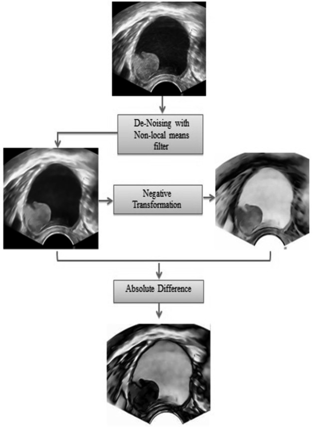
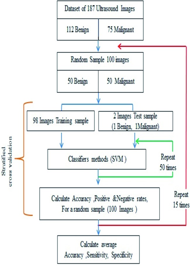
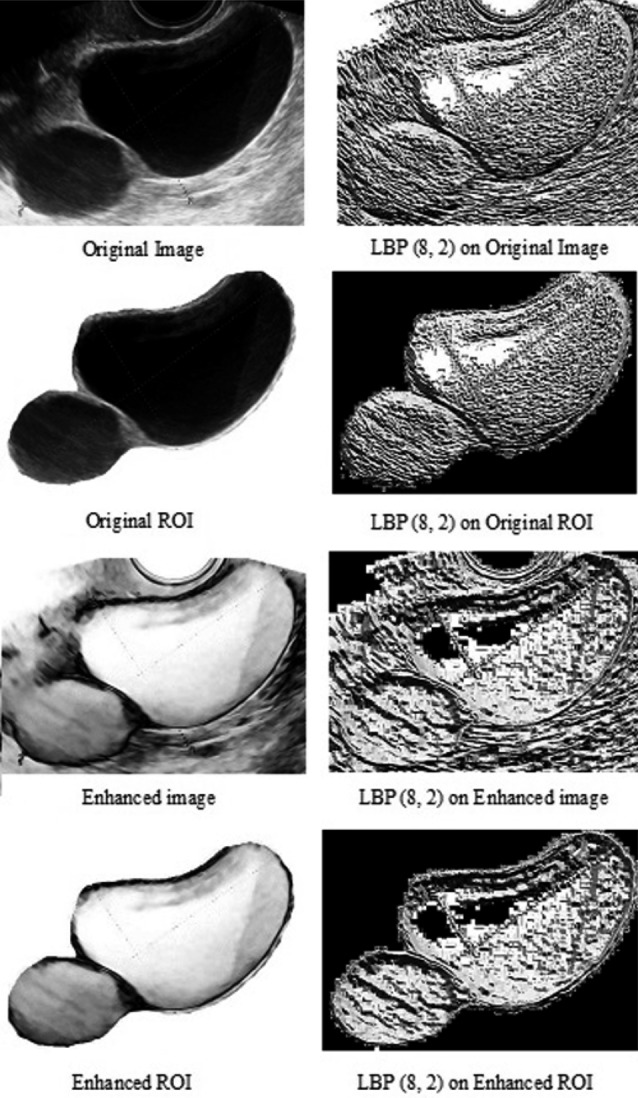
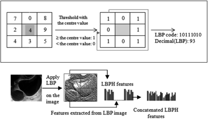
Transvaginal 2D B mode static ultrasound images of 187 ovarian masses with known histological diagnosis were included. Images were first pre-processed and enhanced, and Local Binary Pattern Histograms were then extracted from 2 × 2 blocks of each image. A Support Vector Machine (SVM) was trained using stratified cross validation with randomised sampling. The process was repeated 15 times and in each round 100 images were randomly selected.

RESULTS

The SVM classified the original non-treated static images as benign or malignant masses with an average accuracy of 0.62 (95% CI: 0.59-0.65). This performance significantly improved to an average accuracy of 0.77 (95% CI: 0.75-0.79) when images were pre-processed, enhanced and treated with a Local Binary Pattern operator (mean difference 0.15: 95% 0.11-0.19, p < 0.0001, two-tailed t test).

CONCLUSION

We have shown that an SVM can classify static 2D B mode ultrasound images of ovarian masses into benign and malignant categories. The accuracy improves if texture related LBP features extracted from the images are considered.

摘要

引言

术前将卵巢肿块区分为良性或恶性对于优化患者管理至关重要。

目的

在本研究中,我们开发并验证了一种用于将卵巢肿块区分为良性或恶性的计算机模型。

材料与方法

纳入了187个具有已知组织学诊断的卵巢肿块的经阴道二维B模式静态超声图像。图像首先进行预处理和增强,然后从每个图像的2×2块中提取局部二值模式直方图。使用分层交叉验证和随机抽样对支持向量机(SVM)进行训练。该过程重复15次,每轮随机选择100张图像。

结果

SVM将原始未处理的静态图像分类为良性或恶性肿块,平均准确率为0.62(95%可信区间:0.59 - 0.65)。当图像进行预处理、增强并使用局部二值模式算子处理后,该性能显著提高至平均准确率0.77(95%可信区间:0.75 - 0.79)(平均差异0.15:95% 0.11 - 0.19,p < 0.0001,双尾t检验)。

结论

我们已经表明,支持向量机可以将卵巢肿块的静态二维B模式超声图像分类为良性和恶性类别。如果考虑从图像中提取的与纹理相关的局部二值模式特征,准确率会提高。

https://cdn.ncbi.nlm.nih.gov/pmc/blobs/6b13/4402446/5b398e1fdb5c/FVVinObGyn-7-7-15-g005.jpg
https://cdn.ncbi.nlm.nih.gov/pmc/blobs/6b13/4402446/0fed01eda6ab/FVVinObGyn-7-7-15-g001.jpg
https://cdn.ncbi.nlm.nih.gov/pmc/blobs/6b13/4402446/fd02d68a3f2a/FVVinObGyn-7-7-15-g002.jpg
https://cdn.ncbi.nlm.nih.gov/pmc/blobs/6b13/4402446/57a2fd022500/FVVinObGyn-7-7-15-g003.jpg
https://cdn.ncbi.nlm.nih.gov/pmc/blobs/6b13/4402446/640df1cf87cc/FVVinObGyn-7-7-15-g004.jpg
https://cdn.ncbi.nlm.nih.gov/pmc/blobs/6b13/4402446/5b398e1fdb5c/FVVinObGyn-7-7-15-g005.jpg

相似文献

1
Automated characterisation of ultrasound images of ovarian tumours: the diagnostic accuracy of a support vector machine and image processing with a local binary pattern operator.卵巢肿瘤超声图像的自动特征提取:支持向量机与局部二值模式算子图像处理的诊断准确性
Facts Views Vis Obgyn. 2015;7(1):7-15.
2
An Evaluation of Effectiveness of a Texture Feature Based Computerized Diagnostic Model in Classifying the Ovarian Cyst as Benign and Malignant from Static 2D B-Mode Ultrasound Images.基于纹理特征的计算机诊断模型对静态 2D 灰阶超声图像中卵巢囊肿良恶性分类的有效性评估。
Curr Med Imaging. 2023;19(3):292-305. doi: 10.2174/1573405618666220516120556.
3
An Evaluation of the Effectiveness of Image-based Texture Features Extracted from Static B-mode Ultrasound Images in Distinguishing between Benign and Malignant Ovarian Masses.基于静态 B 型超声图像提取的纹理特征在鉴别良恶性卵巢肿块中的有效性评估。
Ultrason Imaging. 2021 May;43(3):124-138. doi: 10.1177/0161734621998091. Epub 2021 Feb 25.
4
Differentiation of fat-poor angiomyolipoma from clear cell renal cell carcinoma in contrast-enhanced MDCT images using quantitative feature classification.基于定量特征分类的 MDCT 增强图像鉴别乏脂性血管平滑肌脂肪瘤与透明细胞肾细胞癌
Med Phys. 2017 Jul;44(7):3604-3614. doi: 10.1002/mp.12258. Epub 2017 Jun 9.
5
Non-invasive automated 3D thyroid lesion classification in ultrasound: a class of ThyroScan™ systems.超声下非侵入式自动化 3D 甲状腺病变分类:一类 ThyroScan™ 系统。
Ultrasonics. 2012 Apr;52(4):508-20. doi: 10.1016/j.ultras.2011.11.003. Epub 2011 Nov 25.
6
Evaluation of machine learning methods with Fourier Transform features for classifying ovarian tumors based on ultrasound images.基于超声图像的卵巢肿瘤分类的傅里叶变换特征的机器学习方法评估。
PLoS One. 2019 Jul 26;14(7):e0219388. doi: 10.1371/journal.pone.0219388. eCollection 2019.
7
Ovarian tumor characterization using 3D ultrasound.使用 3D 超声对卵巢肿瘤进行特征描述。
Technol Cancer Res Treat. 2012 Dec;11(6):543-52. doi: 10.7785/tcrt.2012.500272. Epub 2012 Jul 10.
8
Semiquantitative dynamic contrast-enhanced MRI for accurate classification of complex adnexal masses.用于复杂附件包块准确分类的半定量动态对比增强磁共振成像
J Magn Reson Imaging. 2017 Feb;45(2):418-427. doi: 10.1002/jmri.25359. Epub 2016 Jul 1.
9
Patch-based classification of thyroid nodules in ultrasound images using direction independent features extracted by two-threshold binary decomposition.基于补丁的甲状腺结节超声图像分类,使用双阈值二值分解提取的方向无关特征。
Comput Med Imaging Graph. 2019 Jan;71:9-18. doi: 10.1016/j.compmedimag.2018.10.001. Epub 2018 Oct 31.
10
Performance of machine learning software to classify breast lesions using BI-RADS radiomic features on ultrasound images.使用超声图像上的BI-RADS放射组学特征对乳腺病变进行分类的机器学习软件的性能。
Eur Radiol Exp. 2019 Aug 5;3(1):34. doi: 10.1186/s41747-019-0112-7.

引用本文的文献

1
AI-based automated segmentation for ovarian/adnexal masses and their internal components on ultrasound imaging.基于人工智能的超声成像中卵巢/附件肿块及其内部成分的自动分割
J Med Imaging (Bellingham). 2024 Jul;11(4):044505. doi: 10.1117/1.JMI.11.4.044505. Epub 2024 Aug 6.
2
Artificial intelligence as a teaching tool for gynaecological ultrasound: A systematic search and scoping review.人工智能作为妇科超声的教学工具:一项系统检索与范围综述
Australas J Ultrasound Med. 2023 Nov 20;27(1):5-11. doi: 10.1002/ajum.12368. eCollection 2024 Feb.
3
Analysis of computer-aided diagnostics in the preoperative diagnosis of ovarian cancer: a systematic review.

本文引用的文献

1
Multicentre external validation of IOTA prediction models and RMI by operators with varied training.多中心验证 IOTA 预测模型和 RMI 对具有不同培训经验的操作者的适用性。
Br J Cancer. 2013 Jun 25;108(12):2448-54. doi: 10.1038/bjc.2013.224. Epub 2013 May 14.
2
Ovarian tumor characterization and classification: a class of GyneScan™ systems.
Annu Int Conf IEEE Eng Med Biol Soc. 2012;2012:4446-9. doi: 10.1109/EMBC.2012.6346953.
3
Evolutionary algorithm-based classifier parameter tuning for automatic ovarian cancer tissue characterization and classification.基于进化算法的分类器参数调整用于卵巢癌组织的自动特征提取与分类
计算机辅助诊断在卵巢癌术前诊断中的分析:一项系统综述。
Insights Imaging. 2023 Feb 15;14(1):34. doi: 10.1186/s13244-022-01345-x.
4
Automatic Detection and Segmentation of Ovarian Cancer Using a Multitask Model in Pelvic CT Images.基于盆腔 CT 图像的多任务模型自动检测和分割卵巢癌。
Oxid Med Cell Longev. 2022 Oct 11;2022:6009107. doi: 10.1155/2022/6009107. eCollection 2022.
5
A machine learning approach applied to gynecological ultrasound to predict progression-free survival in ovarian cancer patients.机器学习方法在妇科超声中的应用,预测卵巢癌患者无进展生存期。
Arch Gynecol Obstet. 2022 Dec;306(6):2143-2154. doi: 10.1007/s00404-022-06578-1. Epub 2022 May 9.
6
The Use of Artificial Intelligence in Automation in the Fields of Gynaecology and Obstetrics - an Assessment of the State of Play.人工智能在妇产科自动化领域的应用——现状评估
Geburtshilfe Frauenheilkd. 2021 Nov 4;81(11):1203-1216. doi: 10.1055/a-1522-3029. eCollection 2021 Nov.
7
A decision support system based on radiomics and machine learning to predict the risk of malignancy of ovarian masses from transvaginal ultrasonography and serum CA-125.基于放射组学和机器学习的决策支持系统,用于从经阴道超声和血清 CA-125 预测卵巢肿块的恶性风险。
Eur Radiol Exp. 2021 Jul 26;5(1):28. doi: 10.1186/s41747-021-00226-0.
8
Ultrasonography in the Differentiation of Endometriomas from Hemorrhagic Ovarian Cysts: The Role of Texture Analysis.超声检查在子宫内膜异位囊肿与出血性卵巢囊肿鉴别诊断中的应用:纹理分析的作用
J Pers Med. 2021 Jun 28;11(7):611. doi: 10.3390/jpm11070611.
9
Ultrasonography in the Diagnosis of Adnexal Lesions: The Role of Texture Analysis.超声检查在附件区病变诊断中的应用:纹理分析的作用
Diagnostics (Basel). 2021 Apr 29;11(5):812. doi: 10.3390/diagnostics11050812.
10
Technology trends and applications of deep learning in ultrasonography: image quality enhancement, diagnostic support, and improving workflow efficiency.深度学习在超声检查中的技术趋势与应用:图像质量增强、诊断支持及工作流程效率提升
Ultrasonography. 2021 Jan;40(1):7-22. doi: 10.14366/usg.20102. Epub 2020 Sep 14.
Ultraschall Med. 2014 Jun;35(3):237-45. doi: 10.1055/s-0032-1330336. Epub 2012 Dec 20.
4
Improving strategies for diagnosing ovarian cancer: a summary of the International Ovarian Tumor Analysis (IOTA) studies.改善卵巢癌诊断策略:国际卵巢肿瘤分析(IOTA)研究总结。
Ultrasound Obstet Gynecol. 2013 Jan;41(1):9-20. doi: 10.1002/uog.12323.
5
Simple ultrasound rules to distinguish between benign and malignant adnexal masses before surgery: prospective validation by IOTA group.术前简单超声规则区分附件良恶性肿块:IOTA 组的前瞻性验证。
BMJ. 2010 Dec 14;341:c6839. doi: 10.1136/bmj.c6839.
6
Ovarian cancer prediction in adnexal masses using ultrasound-based logistic regression models: a temporal and external validation study by the IOTA group.基于超声的逻辑回归模型对附件包块进行卵巢癌预测:IOTA 小组的时间和外部验证研究。
Ultrasound Obstet Gynecol. 2010 Aug;36(2):226-34. doi: 10.1002/uog.7636.
7
Real-time ultrasound vs. evaluation of static images in the preoperative assessment of adnexal masses.实时超声与静态图像评估在附件包块术前评估中的比较
Ultrasound Obstet Gynecol. 2008 Nov;32(6):828-31. doi: 10.1002/uog.6214.
8
Preoperative diagnosis of ovarian tumors using Bayesian kernel-based methods.基于贝叶斯核方法的卵巢肿瘤术前诊断
Ultrasound Obstet Gynecol. 2007 May;29(5):496-504. doi: 10.1002/uog.3996.
9
Texture analysis of medical images.医学图像的纹理分析
Clin Radiol. 2004 Dec;59(12):1061-9. doi: 10.1016/j.crad.2004.07.008.
10
An automatic approach for morphological analysis and malignancy evaluation of ovarian masses using B-scans.
Ultrasound Med Biol. 2003 Nov;29(11):1561-70. doi: 10.1016/j.ultrasmedbio.2003.08.013.