Wong Man Yan, Cavolo Samantha L, Levitan Edwin S
Department of Pharmacology and Chemical Biology, University of Pittsburgh School of Medicine, Pittsburgh, PA 15261.
Department of Pharmacology and Chemical Biology, University of Pittsburgh School of Medicine, Pittsburgh, PA 15261
Mol Biol Cell. 2015 Jul 1;26(13):2466-74. doi: 10.1091/mbc.E15-01-0002. Epub 2015 Apr 22.
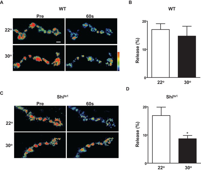
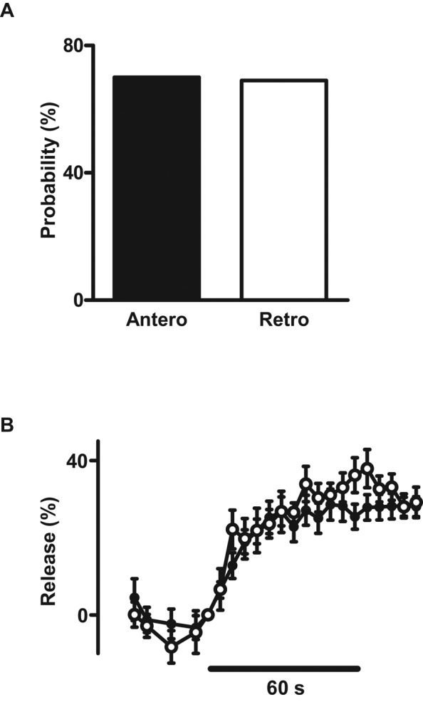
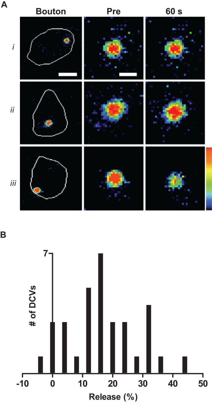
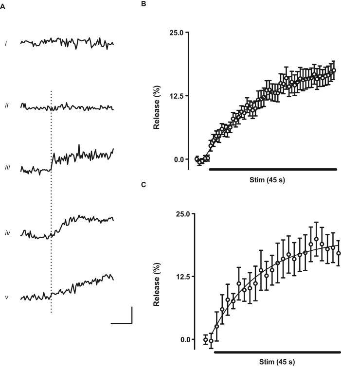
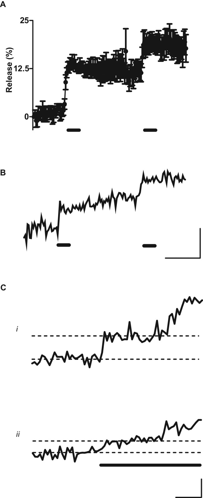
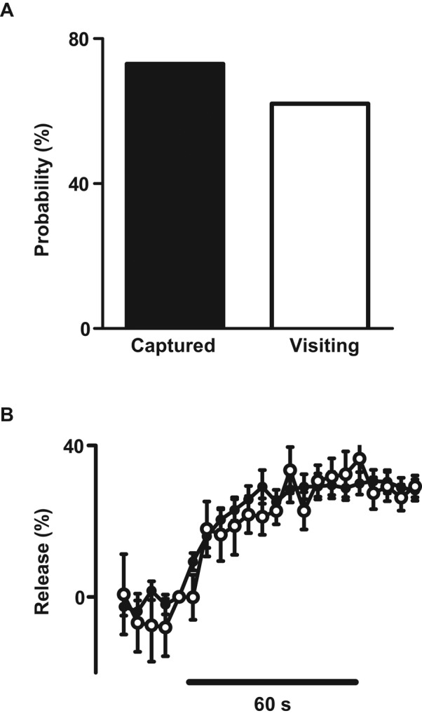
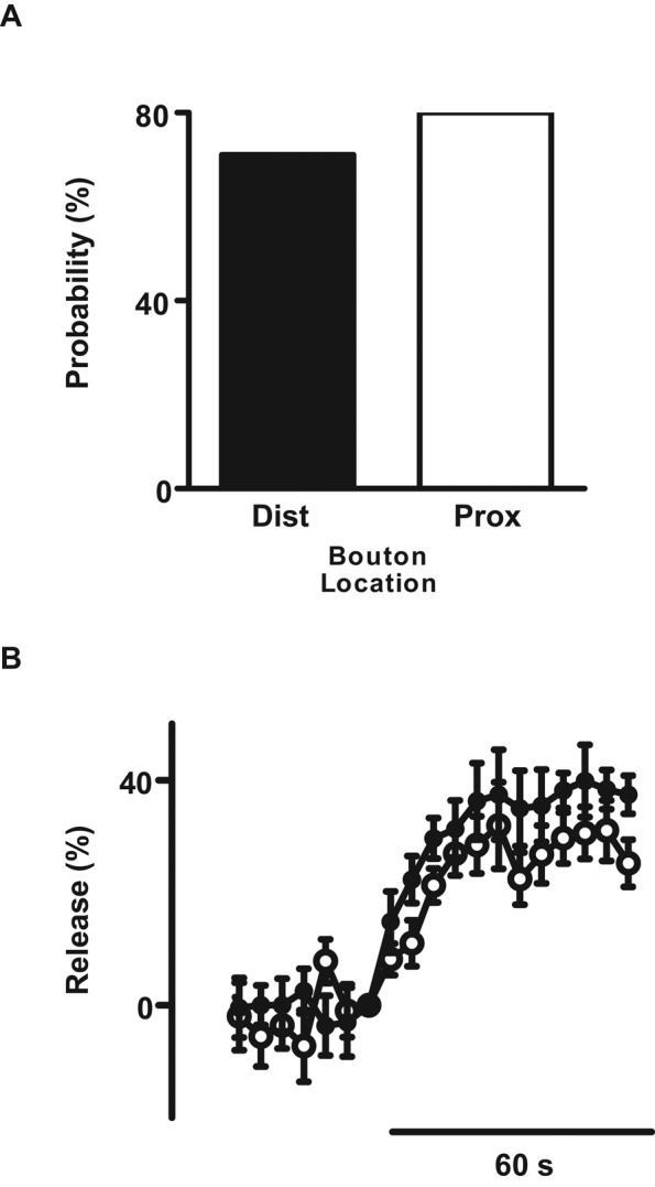
Neurons release neuropeptides, enzymes, and neurotrophins by exocytosis of dense-core vesicles (DCVs). Peptide release from individual DCVs has been imaged in vitro with endocrine cells and at the neuron soma, growth cones, neurites, axons, and dendrites but not at nerve terminals, where peptidergic neurotransmission occurs. Single presynaptic DCVs have, however, been tracked in native terminals with simultaneous photobleaching and imaging (SPAIM) to show that DCVs undergo anterograde and retrograde capture as they circulate through en passant boutons. Here dynamin (encoded by the shibire gene) is shown to enhance activity-evoked peptide release at the Drosophila neuromuscular junction. SPAIM demonstrates that activity depletes only a portion of a single presynaptic DCV's content. Activity initiates exocytosis within seconds, but subsequent release occurs slowly. Synaptic neuropeptide release is further sustained by DCVs undergoing multiple rounds of exocytosis. Synaptic neuropeptide release is surprisingly similar regardless of anterograde or retrograde DCV transport into boutons, bouton location, and time of arrival in the terminal. Thus vesicle circulation and bidirectional capture supply synapses with functionally competent DCVs. These results show that activity-evoked synaptic neuropeptide release is independent of a DCV's past traffic and occurs by slow, dynamin-dependent partial emptying of DCVs, suggestive of kiss-and-run exocytosis.
神经元通过致密核心囊泡(DCV)的胞吐作用释放神经肽、酶和神经营养因子。单个DCV的肽释放已在体外利用内分泌细胞以及在神经元胞体、生长锥、神经突、轴突和树突中成像,但尚未在发生肽能神经传递的神经末梢成像。然而,已利用同步光漂白和成像(SPAIM)在天然末梢中追踪单个突触前DCV,以表明DCV在通过旁触体循环时经历顺行和逆行捕获。在这里,发动蛋白(由发动蛋白基因编码)被证明可增强果蝇神经肌肉接头处活动诱发的肽释放。SPAIM表明,活动仅耗尽单个突触前DCV一部分内容物。活动在数秒内引发胞吐作用,但随后的释放发生缓慢。突触神经肽的释放通过经历多轮胞吐作用的DCV进一步维持。无论DCV是顺行还是逆行转运到旁触体、旁触体位置以及到达末梢的时间如何,突触神经肽释放都惊人地相似。因此,囊泡循环和双向捕获为突触提供功能上正常的DCV。这些结果表明,活动诱发的突触神经肽释放独立于DCV过去的运输过程,并且通过发动蛋白依赖的DCV缓慢部分排空而发生,提示为亲吻-逃离式胞吐作用。