Li Ling-Jun, Aris Izzuddin, Su Lin Lin, Tint Mya Thway, Cheung Carol Yim-Lui, Ikram M Kamran, Gluckman Peter, Godfrey Keith M, Tan Kok Hian, Yeo George, Yap Fabian, Kwek Kenneth, Saw Seang-Mei, Chong Yap-Seng, Wong Tien-Yin, Lee Yung Seng
Singapore Eye Research Institute, Singapore National Eye Centre, Singapore.
Department of Pediatrics, Yong Loo Lin School of Medicine, National University of Singapore, Singapore.
PLoS One. 2015 Apr 24;10(4):e0118250. doi: 10.1371/journal.pone.0118250. eCollection 2015.
We aimed to study the maternal retinal microvasculature at mid-trimester and its relationship with subsequent fetal growth and birth size.
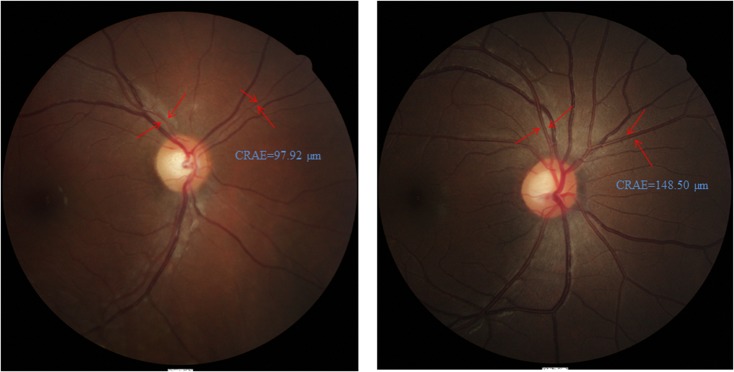
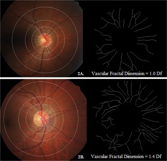
We recruited 732 pregnant women aged 18-46 years in the first trimester with singleton pregnancies. All had retinal photography and fetal scan performed at 26-28 weeks gestation, and subsequent fetal scan at 32-34 weeks gestation. Infant anthropometric measurements were done at birth. Retinal microvasculature was measured using computer software from the retinal photographs.
In multiple linear regression models, each 10 μm narrowing in maternal retinal arteriolar caliber was associated with decreases of 1.36 mm in fetal head circumference at 32-34 weeks gestation, as well as decreases of 1.50 mm and 2.30 mm in infant head circumference and birth length at delivery, respectively. Each standard deviation decrease in maternal retinal arteriolar fractal dimension was associated with decreases of 1.55 mm in fetal head circumference at 32-34 weeks gestation, as well as decreases of 1.08 mm and 46.42 g in infant head circumference and birth weight at delivery, respectively.
Narrower retinal arteriolar caliber and a sparser retinal vascular network in mothers, reflecting a suboptimal uteroplacental microvasculature during mid-pregnancy, were associated with poorer fetal growth and birth size.
我们旨在研究孕中期孕妇的视网膜微血管系统及其与随后胎儿生长和出生体重的关系。
我们招募了732名年龄在18 - 46岁之间、孕早期为单胎妊娠的孕妇。所有孕妇在妊娠26 - 28周时进行了视网膜摄影和胎儿扫描,并在妊娠32 - 34周时进行了后续胎儿扫描。婴儿出生时进行了人体测量。使用计算机软件从视网膜照片中测量视网膜微血管系统。
在多元线性回归模型中,孕妇视网膜小动脉管径每变窄10μm,与妊娠32 - 34周时胎儿头围减少1.36mm相关,同时与婴儿出生时头围减少1.50mm和出生身长减少2.30mm相关。孕妇视网膜小动脉分形维数每降低一个标准差,与妊娠32 - 34周时胎儿头围减少1.55mm相关,同时与婴儿出生时头围减少1.08mm和出生体重减少46.42g相关。
母亲视网膜小动脉管径变窄和视网膜血管网络稀疏,反映了妊娠中期子宫胎盘微血管系统欠佳,与胎儿生长和出生体重较差有关。