Tiwari Saumya, Reddy Vijaya B, Bhargava Rohit, Raman Jaishankar
Department of Bioengineering, Beckman Institute for Advanced Science and Technology, University of Illinois at Urbana Champaign, Urbana, Illinois, 61801, United States of America.
Department of Pathology, Rush University Medical Center, 1725 West Harrison St, Chicago, Illinois, 60612, United States of America.
PLoS One. 2015 May 1;10(5):e0125183. doi: 10.1371/journal.pone.0125183. eCollection 2015.
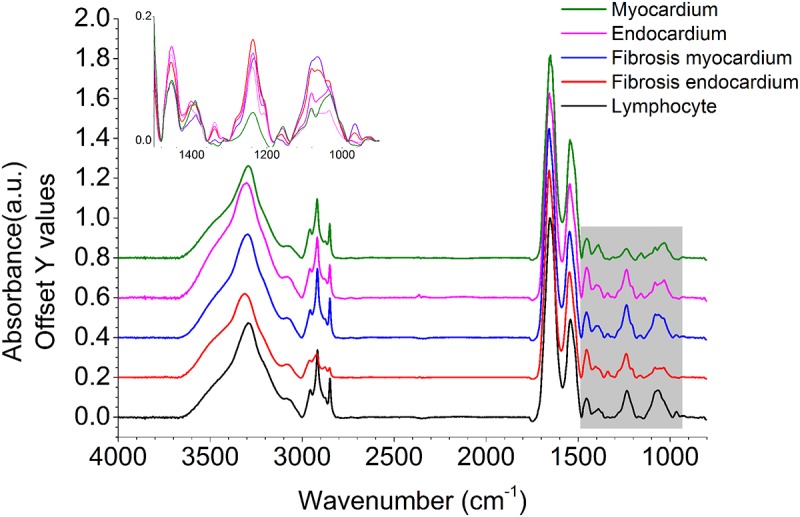
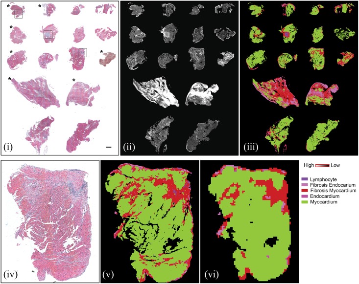
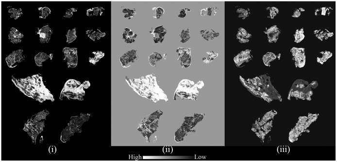
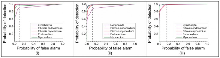
Rejection is a common problem after cardiac transplants leading to significant number of adverse events and deaths, particularly in the first year of transplantation. The gold standard to identify rejection is endomyocardial biopsy. This technique is complex, cumbersome and requires a lot of expertise in the correct interpretation of stained biopsy sections. Traditional histopathology cannot be used actively or quickly during cardiac interventions or surgery. Our objective was to develop a stain-less approach using an emerging technology, Fourier transform infrared (FT-IR) spectroscopic imaging to identify different components of cardiac tissue by their chemical and molecular basis aided by computer recognition, rather than by visual examination using optical microscopy. We studied this technique in assessment of cardiac transplant rejection to evaluate efficacy in an example of complex cardiovascular pathology. We recorded data from human cardiac transplant patients' biopsies, used a Bayesian classification protocol and developed a visualization scheme to observe chemical differences without the need of stains or human supervision. Using receiver operating characteristic curves, we observed probabilities of detection greater than 95% for four out of five histological classes at 10% probability of false alarm at the cellular level while correctly identifying samples with the hallmarks of the immune response in all cases. The efficacy of manual examination can be significantly increased by observing the inherent biochemical changes in tissues, which enables us to achieve greater diagnostic confidence in an automated, label-free manner. We developed a computational pathology system that gives high contrast images and seems superior to traditional staining procedures. This study is a prelude to the development of real time in situ imaging systems, which can assist interventionists and surgeons actively during procedures.
排斥反应是心脏移植后常见的问题,会导致大量不良事件和死亡,尤其是在移植后的第一年。识别排斥反应的金标准是心内膜心肌活检。这项技术复杂、繁琐,并且在正确解读染色活检切片方面需要很多专业知识。在心脏介入手术或外科手术过程中,传统组织病理学无法快速有效地发挥作用。我们的目标是开发一种无需染色的方法,利用新兴技术傅里叶变换红外(FT-IR)光谱成像,借助计算机识别,根据化学和分子基础来识别心脏组织的不同成分,而不是通过光学显微镜的目视检查。我们在心脏移植排斥反应的评估中研究了这项技术,以复杂心血管病理为例评估其有效性。我们记录了人类心脏移植患者活检的数据,使用贝叶斯分类方案并开发了一种可视化方案,无需染色或人工监督即可观察化学差异。使用受试者工作特征曲线,我们发现在细胞水平上误报概率为10%时,五种组织学类别中有四种的检测概率大于95%,并且在所有情况下都能正确识别具有免疫反应特征的样本。通过观察组织中固有的生化变化,可以显著提高人工检查的效率,这使我们能够以自动化、无标记的方式获得更高的诊断置信度。我们开发了一种计算病理学系统,该系统能提供高对比度图像,似乎优于传统染色程序。这项研究是实时原位成像系统开发的前奏,该系统可以在手术过程中积极协助介入医生和外科医生。