• 文献检索
  • 文档翻译
  • 深度研究
  • 学术资讯
  • Suppr Zotero 插件Zotero 插件
  • 邀请有礼
  • 套餐&价格
  • 历史记录
应用&插件
Suppr Zotero 插件Zotero 插件浏览器插件Mac 客户端Windows 客户端微信小程序
定价
高级版会员购买积分包购买API积分包
服务
文献检索文档翻译深度研究API 文档MCP 服务
关于我们
关于 Suppr公司介绍联系我们用户协议隐私条款
关注我们

Suppr 超能文献

核心技术专利:CN118964589B侵权必究
粤ICP备2023148730 号-1Suppr @ 2026

文献检索

告别复杂PubMed语法,用中文像聊天一样搜索,搜遍4000万医学文献。AI智能推荐,让科研检索更轻松。

立即免费搜索

文件翻译

保留排版,准确专业,支持PDF/Word/PPT等文件格式,支持 12+语言互译。

免费翻译文档

深度研究

AI帮你快速写综述,25分钟生成高质量综述,智能提取关键信息,辅助科研写作。

立即免费体验

扩散加权磁共振神经成像在肿瘤学实践中用于评估臂丛神经病变

Diffusion-weighted MR neurography for the assessment of brachial plexopathy in oncological practice.

作者信息

Andreou Adrian, Sohaib Aslam, Collins David J, Takahara Taro, Kwee Thomas C, Leach Martin O, MacVicar David A, Koh Dow-Mu

机构信息

Department of Radiology, Royal Marsden, Sutton, Surrey, UK.

CR UK Clinical Magnetic Resonance Research Group, Institute of Cancer Research, Sutton, Surrey, UK.

出版信息

Cancer Imaging. 2015 May 2;15(1):6. doi: 10.1186/s40644-015-0041-5.

DOI:10.1186/s40644-015-0041-5
PMID:25934632
原文链接:https://pmc.ncbi.nlm.nih.gov/articles/PMC4417248/
Abstract

BACKGROUND

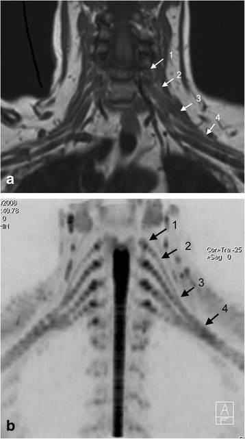
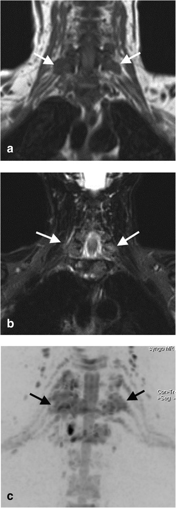
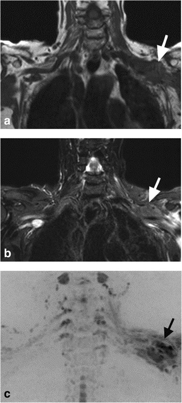
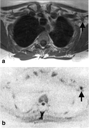
To evaluate diffusion-weighted MR neurography (DW-MRN) for visualizing the brachial plexus and for the assessment of brachial plexopathy.

METHODS

40 oncological patients with symptoms of brachial plexopathy underwent 1.5 T MRI using conventional MR sequences and unidirectional DW-MRN. The images were independently reviewed by two radiologists. Anatomic visualization of the brachial plexus was scored using a 5 point scale on conventional MR sequences and then combined with DW-MRN. A brachial plexus abnormality was also scored using a 5 point scale and inter-observer agreement determined by kappa statistics. Diagnostic accuracy for brachial plexopathy assessed by conventional MRI alone versus conventional MRI combined with DW-MRN was compared by ROC analysis using reference standards.

RESULTS

DW-MRN significantly improved visualization of the brachial plexus compared with conventional MRI alone (P<0.001). When assessing brachial plexopathy, inter-observer agreement was moderate for conventional MRI (kappa=0.48) but good for conventional MRI with DW-MRN (kappa=0.62). DW-MRN combined with conventional MRI significantly improved diagnostic accuracy in one observer (P<0.05) but was similar in the other observer.

CONCLUSION

DW-MRN improved visualization of the brachial plexus. Combining DW-MRN with conventional MRI can improve inter-observer agreement and detection of brachial plexopathy in symptomatic oncological patients.

摘要

背景

评估扩散加权磁共振神经成像(DW-MRN)用于可视化臂丛神经及评估臂丛神经病变的情况。

方法

40例有臂丛神经病变症状的肿瘤患者接受了1.5T磁共振成像检查,使用传统磁共振序列和单向DW-MRN。图像由两名放射科医生独立评估。在传统磁共振序列上使用5分制对臂丛神经的解剖可视化进行评分,然后与DW-MRN相结合。臂丛神经异常也使用5分制进行评分,并通过kappa统计确定观察者间的一致性。使用参考标准通过ROC分析比较单独使用传统MRI与传统MRI联合DW-MRN评估臂丛神经病变的诊断准确性。

结果

与单独使用传统MRI相比,DW-MRN显著改善了臂丛神经的可视化(P<0.001)。在评估臂丛神经病变时,传统MRI的观察者间一致性为中等(kappa=0.48),而传统MRI联合DW-MRN时为良好(kappa=0.62)。DW-MRN与传统MRI联合使用在一名观察者中显著提高了诊断准确性(P<0.05),但在另一名观察者中相似。

结论

DW-MRN改善了臂丛神经的可视化。将DW-MRN与传统MRI相结合可以提高有症状肿瘤患者中观察者间的一致性以及臂丛神经病变的检测率。

https://cdn.ncbi.nlm.nih.gov/pmc/blobs/3300/4417248/c9e37cff2a24/40644_2015_41_Fig4_HTML.jpg
https://cdn.ncbi.nlm.nih.gov/pmc/blobs/3300/4417248/68c0606d4a2c/40644_2015_41_Fig1_HTML.jpg
https://cdn.ncbi.nlm.nih.gov/pmc/blobs/3300/4417248/437768330b38/40644_2015_41_Fig2_HTML.jpg
https://cdn.ncbi.nlm.nih.gov/pmc/blobs/3300/4417248/0b67b846a5c3/40644_2015_41_Fig3_HTML.jpg
https://cdn.ncbi.nlm.nih.gov/pmc/blobs/3300/4417248/c9e37cff2a24/40644_2015_41_Fig4_HTML.jpg

相似文献

1
Diffusion-weighted MR neurography for the assessment of brachial plexopathy in oncological practice.扩散加权磁共振神经成像在肿瘤学实践中用于评估臂丛神经病变
Cancer Imaging. 2015 May 2;15(1):6. doi: 10.1186/s40644-015-0041-5.
2
Accuracy of MR neurography in the diagnosis of brachial plexopathy.磁共振神经成像在臂丛神经病诊断中的准确性。
Eur J Radiol. 2017 Oct;95:24-27. doi: 10.1016/j.ejrad.2017.07.012. Epub 2017 Jul 19.
3
Diagnostic performance of diffusion-weighted MR neurography as an adjunct to conventional MRI for the assessment of brachial plexus pathology.扩散加权磁共振神经造影作为传统MRI的辅助手段用于评估臂丛神经病变的诊断效能。
Eur Radiol. 2022 Apr;32(4):2791-2797. doi: 10.1007/s00330-021-08324-8. Epub 2021 Nov 9.
4
Contrast-enhanced magnetic resonance neurography for diagnosing brachial plexopathy: improved visualization and additional imaging features.对比增强磁共振神经成像在诊断臂丛神经病中的应用:改善可视化和附加成像特征。
Sci Rep. 2024 Sep 5;14(1):20758. doi: 10.1038/s41598-024-71554-5.
5
Diffusion-weighted MR neurography of median and ulnar nerves in the wrist and palm.手腕和手掌部正中神经与尺神经的磁共振神经造影弥散加权成像
Eur Radiol. 2017 Jun;27(6):2359-2366. doi: 10.1007/s00330-016-4591-0. Epub 2016 Sep 15.
6
MR neurography in traumatic brachial plexopathy.创伤性臂丛神经病的磁共振神经成像
Eur J Radiol. 2015 May;84(5):927-32. doi: 10.1016/j.ejrad.2015.02.006. Epub 2015 Feb 16.
7
Diagnostic Value and Surgical Implications of the 3D DW-SSFP MRI On the Management of Patients with Brachial Plexus Injuries.三维DW-SSFP MRI在臂丛神经损伤患者管理中的诊断价值及手术意义
Sci Rep. 2016 Oct 26;6:35999. doi: 10.1038/srep35999.
8
Combine contrast-enhanced 3D T2-weighted short inversion time inversion recovery MR neurography with MR angiography at 1.5 T in the assessment of brachial plexopathy.在 1.5T 场强下,结合对比增强三维 T2 加权短反转时间反转恢复磁共振神经成像和磁共振血管成像评估臂丛神经病。
MAGMA. 2021 Apr;34(2):229-239. doi: 10.1007/s10334-020-00867-z. Epub 2020 Jul 13.
9
Three-tesla magnetic resonance neurography of the brachial plexus in cervical radiculopathy.用于诊断神经根型颈椎病的臂丛神经3特斯拉磁共振神经成像
Muscle Nerve. 2015 Sep;52(3):392-6. doi: 10.1002/mus.24560. Epub 2015 Jun 18.
10
Selective visualization of pelvic splanchnic nerve and pelvic plexus using readout-segmented echo-planar diffusion-weighted magnetic resonance neurography: A preliminary study in healthy male volunteers.利用读出分段回波平面扩散加权磁共振神经成像对盆内脏神经和盆腔神经丛进行选择性可视化:一项在健康男性志愿者中的初步研究。
Eur J Radiol. 2017 Jan;86:52-57. doi: 10.1016/j.ejrad.2016.10.034. Epub 2016 Nov 2.

引用本文的文献

1
Diffusion-Weighted MR Neurography with Unidirectional Motion-Probing Gradient to Evaluate Lumbar Nerve Roots at 1.5T MR.利用单向运动探测梯度的扩散加权磁共振神经成像在1.5T磁共振成像上评估腰神经根
J Korean Soc Radiol. 2024 May;85(3):607-617. doi: 10.3348/jksr.2023.0052. Epub 2024 Mar 8.
2
Clinical Application of Diffusion Tensor Imaging for a Brachial Plexus Injury.扩散张量成像在臂丛神经损伤中的临床应用
Diagnostics (Basel). 2022 Jul 11;12(7):1687. doi: 10.3390/diagnostics12071687.
3
Diagnostic performance of diffusion-weighted MR neurography as an adjunct to conventional MRI for the assessment of brachial plexus pathology.

本文引用的文献

1
3T MR tomography of the brachial plexus: structural and microstructural evaluation.3T 磁共振臂丛神经成像:结构与微观结构评估。
Eur J Radiol. 2012 Sep;81(9):2231-45. doi: 10.1016/j.ejrad.2011.05.021. Epub 2011 Jul 16.
2
New approaches in imaging of the brachial plexus.臂丛神经影像学的新方法。
Eur J Radiol. 2010 May;74(2):403-10. doi: 10.1016/j.ejrad.2010.01.024. Epub 2010 Mar 12.
3
Diffusion-weighted MR neurography of the sacral plexus with unidirectional motion probing gradients.基于单方向运动探测梯度的磁共振弥散加权成像在骶丛神经中的应用。
扩散加权磁共振神经造影作为传统MRI的辅助手段用于评估臂丛神经病变的诊断效能。
Eur Radiol. 2022 Apr;32(4):2791-2797. doi: 10.1007/s00330-021-08324-8. Epub 2021 Nov 9.
4
Multimodality imaging of adult rhabdomyosarcoma: the added value of hybrid imaging.成人横纹肌肉瘤的多模态成像:混合成像的附加价值。
Br J Radiol. 2020 Aug;93(1112):20200250. doi: 10.1259/bjr.20200250. Epub 2020 Jun 26.
5
Diffusion Tensor Imaging of the Brachial Plexus: A Comparison between Readout-segmented and Conventional Single-shot Echo-planar Imaging.臂丛神经的弥散张量成像:分段读出和常规单次激发 echo-planar 成像的比较。
Magn Reson Med Sci. 2019 Apr 10;18(2):150-157. doi: 10.2463/mrms.mp.2018-0004. Epub 2018 Nov 9.
6
Diffusion weighted imaging demystified: the technique and potential clinical applications for soft tissue imaging.弥散加权成像揭秘:软组织成像的技术及潜在临床应用
Skeletal Radiol. 2018 Mar;47(3):313-328. doi: 10.1007/s00256-017-2822-3. Epub 2017 Nov 20.
7
Cystic degeneration of the tibial nerve: magnetic resonance neurography and sonography appearances of an intraneural ganglion cyst.胫神经的囊性退变:神经内腱鞘囊肿的磁共振神经成像和超声表现
Skeletal Radiol. 2017 Dec;46(12):1763-1767. doi: 10.1007/s00256-017-2753-z. Epub 2017 Aug 18.
Eur Radiol. 2010 May;20(5):1221-6. doi: 10.1007/s00330-009-1665-2. Epub 2009 Nov 21.
4
Whole-body magnetic resonance neurography.全身磁共振神经成像
N Engl J Med. 2009 Jul 30;361(5):538-9. doi: 10.1056/NEJMc0902318.
5
Diffusion-weighted MR neurography of the brachial plexus: feasibility study.臂丛神经的扩散加权磁共振神经成像:可行性研究。
Radiology. 2008 Nov;249(2):653-60. doi: 10.1148/radiol.2492071826. Epub 2008 Sep 16.
6
Plexopathy.神经丛病
AJNR Am J Neuroradiol. 2008 Feb;29(2):400-2.
7
MR neurography with multiplanar reconstruction of 3D MRI datasets: an anatomical study and clinical applications.3D MRI数据集多平面重建的磁共振神经成像:解剖学研究与临床应用
Neuroradiology. 2007 Apr;49(4):335-41. doi: 10.1007/s00234-006-0197-6. Epub 2007 Jan 5.
8
Diffusion weighted whole body imaging with background body signal suppression (DWIBS): technical improvement using free breathing, STIR and high resolution 3D display.背景体部信号抑制扩散加权全身成像(DWIBS):利用自由呼吸、短TI反转恢复序列(STIR)及高分辨率三维显示的技术改进
Radiat Med. 2004 Jul-Aug;22(4):275-82.
9
The basis of anisotropic water diffusion in the nervous system - a technical review.神经系统中各向异性水扩散的基础——技术综述
NMR Biomed. 2002 Nov-Dec;15(7-8):435-55. doi: 10.1002/nbm.782.
10
Principles and applications of echo-planar imaging: a review for the general radiologist.回波平面成像的原理与应用:面向普通放射科医生的综述
Radiographics. 2001 May-Jun;21(3):767-79. doi: 10.1148/radiographics.21.3.g01ma23767.