Lymphoma Research Laboratory, Wayne State University School of Medicine, 540 East Canfield, room #8229, Detroit, MI 48202 USA ; Van Elslander Cancer Center, Grosse Pointe Woods, MI USA.
Lymphoma Research Laboratory, Wayne State University School of Medicine, 540 East Canfield, room #8229, Detroit, MI 48202 USA.
Exp Hematol Oncol. 2014 Dec 19;3:31. doi: 10.1186/2162-3619-3-31. eCollection 2014.
The combination of rituximab and 2-CdA is an effective therapy for B-cell tumors. However, the molecular mechanisms and enzymatic pathways involved in the interaction between the two agents are not fully understood. In this study, we provide molecular evidence for positive interaction between these two agents with resultant therapeutic benefit.
Efficacy of the R-2CdA regimen was evaluated in thirteen patients with B-cell tumors (9 CLL; 3 WM and 1 FL), in vitro against 3 lymphoma cell lines and in a xenograft mouse model. Treatment-induced changes involving phenotype, kinase activity and protein expression were assessed in vitro and in the mouse xenograft tumors. The interaction between RTX and 2-CdA was analyzed using the multiple comparison method, Tukey's honestly significant difference (HSD). For the clinical and animal data, survival functions were estimated using the Kaplan-Meier method and compared by the log-rank test. P-values <0.05 were considered statistically significant. All statistical analyses were evaluated using GraphPad Prism 4 (San Diego, CA).
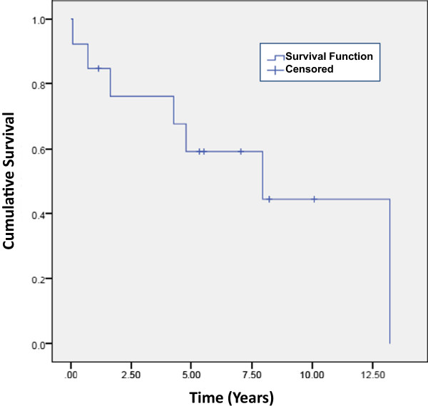
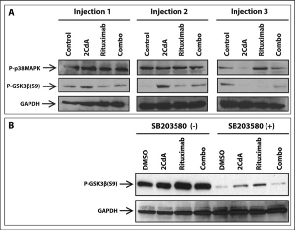
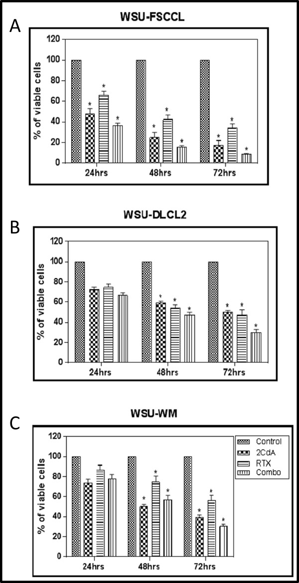
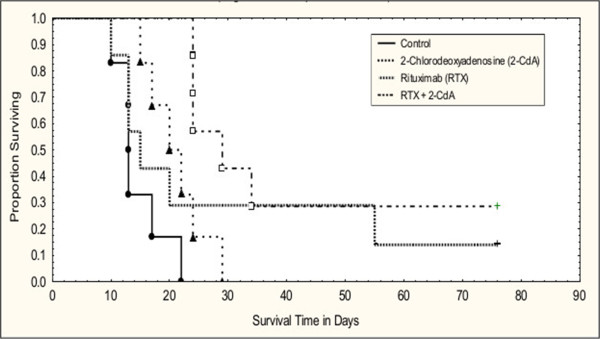
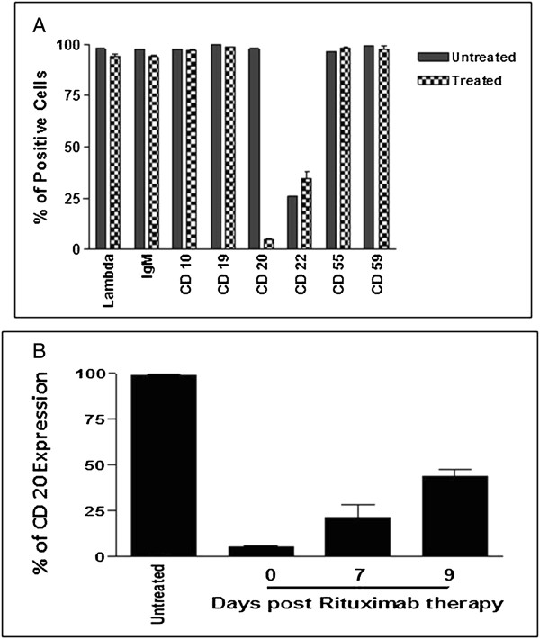
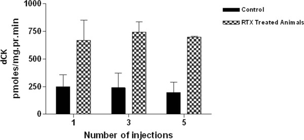
9 of 12 (75%) evaluable patients responded to the R-2-CdA regimen with median duration of response of 34 months. Median survival of patients from diagnosis and from completion of R-2-CdA treatment was 13.3 and 7.9 years, respectively. In vitro, the combination was effective in all 3 cell lines of lymphomas but with higher sensitivity in the follicular lymphoma cell line. The combination was also effective in the WSU-WM-SCID xenograft model with dose-dependent response and synergistic benefit. All animals were tumor-free for up to 120 days post 2 cycles of this regimen. Rituximab induced activation of deoxycytidine kinase (dCK), p38 mitogen activated protein kinase (p38MAPK) and glycogen synthase kinase-3β (GSK-3β) in the xenograft WSU-WM tumors. Chemical inhibition of p38MAPK led to inhibition of the GSK-3β phosphorylation suggesting that GSK-3β is regulated by p38MAPK in this model.
Collectively, our studies show concordance between the activity of R-2-CdA in vitro, in human and in WSU-WM xenograft model attesting to the validity of this model in predicting clinical response. Modulation of dCK and GSK-3β by rituximab may contribute to the positive therapeutic interaction between rituximab and 2-CdA.
利妥昔单抗联合 2-CdA 是治疗 B 细胞肿瘤的有效疗法。然而,这两种药物相互作用涉及的分子机制和酶途径尚未完全了解。在这项研究中,我们提供了分子证据,证明这两种药物具有协同作用,从而带来治疗益处。
我们评估了利妥昔单抗联合 2-CdA(R-2-CdA)方案在 13 例 B 细胞肿瘤患者(9 例 CLL;3 例 WM 和 1 例 FL)中的疗效,在体外针对 3 种淋巴瘤细胞系,并在异种移植小鼠模型中进行了评估。在体外和小鼠异种移植肿瘤中评估了治疗诱导的表型、激酶活性和蛋白表达的变化。使用多重比较法(Tukey 的Honestly Significant Difference(HSD))分析 RTX 和 2-CdA 之间的相互作用。对于临床和动物数据,使用 Kaplan-Meier 方法估计生存函数,并通过对数秩检验进行比较。P 值<0.05 被认为具有统计学意义。所有统计分析均使用 GraphPad Prism 4(圣地亚哥,CA)进行评估。
12 例可评估患者中有 9 例(75%)对 R-2-CdA 方案有反应,中位反应持续时间为 34 个月。患者从诊断到完成 R-2-CdA 治疗的中位生存时间分别为 13.3 年和 7.9 年。在体外,该联合方案对所有 3 种淋巴瘤细胞系均有效,但滤泡性淋巴瘤细胞系的敏感性更高。该联合方案在 WSU-WM-SCID 异种移植模型中也有效,具有剂量依赖性反应和协同作用。所有动物在接受 2 个周期的该方案治疗后,长达 120 天内均未出现肿瘤。利妥昔单抗在异种移植 WSU-WM 肿瘤中诱导脱氧胞苷激酶(dCK)、p38 丝裂原激活蛋白激酶(p38MAPK)和糖原合酶激酶-3β(GSK-3β)的激活。化学抑制 p38MAPK 导致 GSK-3β磷酸化的抑制,表明在该模型中 GSK-3β受 p38MAPK 调节。
综上所述,我们的研究表明 R-2-CdA 在体外、体内和 WSU-WM 异种移植模型中的活性之间具有一致性,证明了该模型在预测临床反应方面的有效性。利妥昔单抗对 dCK 和 GSK-3β 的调节可能有助于利妥昔单抗与 2-CdA 之间的积极治疗相互作用。