Nagai Momoko, Aoyama Tomoki, Ito Akira, Tajino Junichi, Iijima Hirotaka, Yamaguchi Shoki, Zhang Xiangkai, Kuroki Hiroshi
Department of Motor Function Analysis, Human Health Sciences, Graduate School of Medicine, Kyoto University, Kyoto, Japan.
Department of Development and Rehabilitation of Motor Function, Human Health Sciences, Graduate School of Medicine, Kyoto University, Kyoto, Japan.
J Anat. 2015 May;226(5):447-57. doi: 10.1111/joa.12290. Epub 2015 May 4.
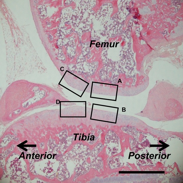
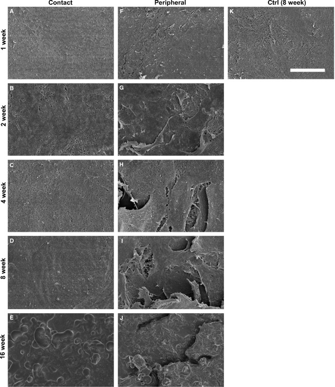
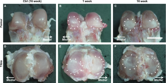
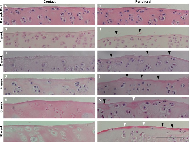
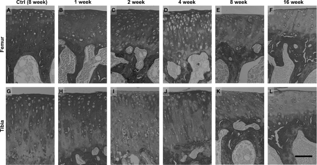
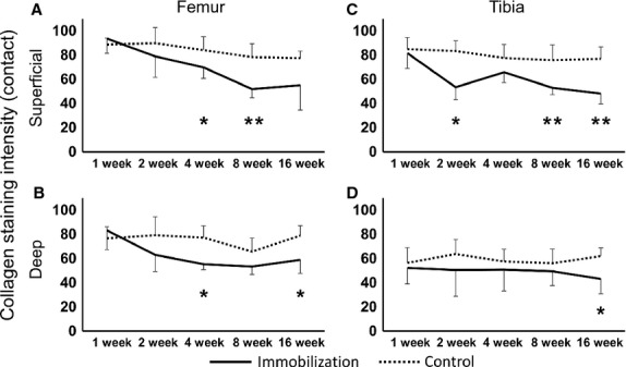
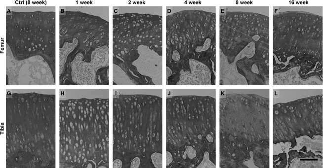
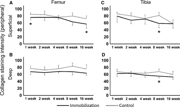
The purpose of this study was to examine the ultrastructural changes of surface cartilage collagen fibers, which differ by region and the length of the experimental period in an immobilization model of rat. Male Wistar rats were randomly divided into histological or macroscopic and ultrastructural assessment groups. The left knees of all the animals were surgically immobilized by external fixation for 1, 2, 4, 8 or 16 weeks (n = 5/time point). Sagittal histological sections of the medial mid-condylar region of the knee were obtained and assessed in four specific regions (contact and peripheral regions of the femur and tibia) and two zones (superficial and deep). To semi-quantify the staining intensity of the collagen fibers in the cartilage, picrosirius red staining was used. The cartilage surface changes of all the assessed regions were investigated by scanning electron microscopy (SEM). From histological and SEM observations, the fibrillation and irregular changes of the cartilage surface were more severe in the peripheral region than in the contact region. Interestingly, at 16 weeks post-immobilization, we observed non-fibrous structures at both the contact and peripheral regions. The collagen fiber staining intensity decreased in the contact region compared with the peripheral region. In conclusion, the alteration of surface collagen fiber ultrastructure and collagen staining intensity differed by the specific cartilage regions after immobilization. These results demonstrate that the progressive degeneration of cartilage is region specific, and depends on the length of the immobilization period.
本研究的目的是在大鼠固定模型中,研究表面软骨胶原纤维的超微结构变化,这些变化因区域和实验周期的长短而异。雄性Wistar大鼠被随机分为组织学或宏观及超微结构评估组。所有动物的左膝均通过外固定手术固定1、2、4、8或16周(每个时间点n = 5)。获取膝关节内侧髁中部区域的矢状组织学切片,并在四个特定区域(股骨和胫骨的接触区和周边区)和两个区域(浅层和深层)进行评估。为了半定量软骨中胶原纤维的染色强度,使用了天狼星红染色。通过扫描电子显微镜(SEM)研究所有评估区域的软骨表面变化。从组织学和SEM观察结果来看,软骨表面的原纤维形成和不规则变化在周边区域比在接触区域更严重。有趣的是,在固定后16周,我们在接触区和周边区均观察到非纤维结构。与周边区域相比,接触区域的胶原纤维染色强度降低。总之,固定后表面胶原纤维超微结构和胶原染色强度的改变因特定软骨区域而异。这些结果表明,软骨的进行性退变具有区域特异性,并取决于固定期的长短。