Quaedflieg C W E M, van de Ven V, Meyer T, Siep N, Merckelbach H, Smeets T
Faculty of Psychology and Neuroscience, Maastricht University, Maastricht, The Netherlands.
PLoS One. 2015 May 6;10(5):e0124141. doi: 10.1371/journal.pone.0124141. eCollection 2015.
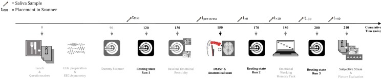
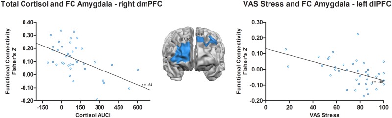
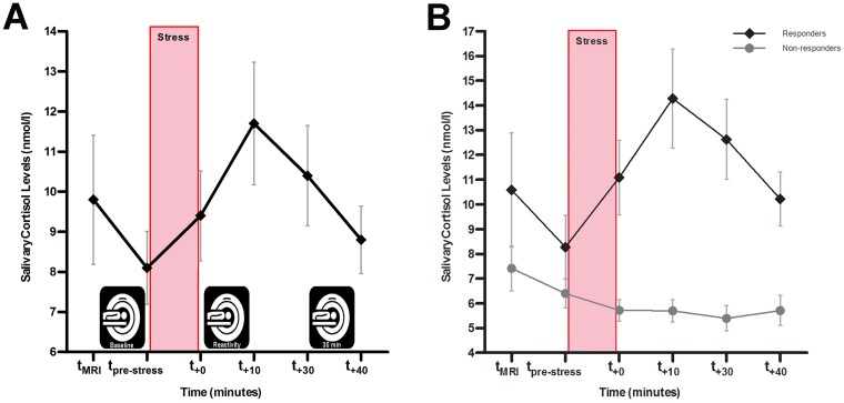
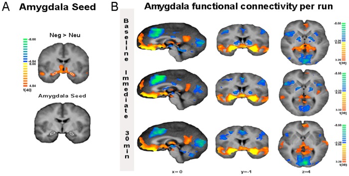
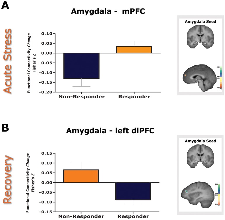
Stress-induced changes in functional brain connectivity have been linked to the etiology of stress-related disorders. Resting state functional connectivity (rsFC) is especially informative in characterizing the temporal trajectory of glucocorticoids during stress adaptation. Using the imaging Maastricht Acute Stress Test (iMAST), we induced acute stress in 39 healthy volunteers and monitored the neuroendocrine stress levels during three runs of resting state functional magnetic resonance imaging (rs-fMRI): before (run 1), immediately following (run 2), and 30 min after acute stress (run 3). The iMAST resulted in strong increases in cortisol levels. Whole-brain analysis revealed that acute stress (run 2 - 1) was characterized by changes in connectivity of the amygdala with the ventrolateral prefrontal cortex (vlPFC), ventral posterior cingulate cortex (PCC), cuneus, parahippocampal gyrus, and culmen. Additionally, cortisol responders were characterized by enhanced amygdala - medial prefrontal cortex (mPFC) connectivity. Stress recovery (run 3 - 2) was characterized by altered amygdala connectivity with the dorsolateral prefrontal cortex (dlPFC), ventral and dorsal anterior cingulate cortex (ACC), anterior hippocampal complex, cuneus, and presupplementary motor area (preSMA). Opposite to non-responders, cortisol responders were characterized by enhanced amygdala connectivity with the anterior hippocampal complex and parahippocampal gyrus, and reduced connectivity with left dlPFC, dACC, and culmen during early recovery. Acute stress responding and recovery are thus associated with changes in the functional connectivity of the amygdala network. Our findings show that these changes may be regulated via stress-induced neuroendocrine levels. Defining stress-induced neuronal network changes is pertinent to developing treatments that target abnormal neuronal activity.
应激诱导的大脑功能连接变化与应激相关障碍的病因有关。静息态功能连接(rsFC)在表征应激适应过程中糖皮质激素的时间轨迹方面尤其具有信息量。使用成像马斯特里赫特急性应激试验(iMAST),我们在39名健康志愿者中诱导急性应激,并在静息态功能磁共振成像(rs-fMRI)的三次扫描过程中监测神经内分泌应激水平:急性应激前(扫描1)、急性应激后立即(扫描2)和急性应激后30分钟(扫描3)。iMAST导致皮质醇水平大幅升高。全脑分析显示,急性应激(扫描2 - 1)的特征是杏仁核与腹外侧前额叶皮质(vlPFC)、腹侧后扣带回皮质(PCC)、楔叶、海马旁回和山顶的连接发生变化。此外,皮质醇反应者的特征是杏仁核与内侧前额叶皮质(mPFC)的连接增强。应激恢复(扫描3 - 2)的特征是杏仁核与背外侧前额叶皮质(dlPFC)、腹侧和背侧前扣带回皮质(ACC)、前海马复合体、楔叶和辅助运动前区(preSMA)的连接改变。与无反应者相反,皮质醇反应者的特征是在早期恢复期间,杏仁核与前海马复合体和海马旁回的连接增强,与左侧dlPFC、背侧ACC和山顶的连接减少。因此,急性应激反应和恢复与杏仁核网络功能连接的变化有关。我们的研究结果表明,这些变化可能通过应激诱导的神经内分泌水平来调节。定义应激诱导的神经元网络变化与开发针对异常神经元活动的治疗方法相关。