Wang Li, Xing Jie, Cheng Rui, Shao Ying, Li Peng, Zhu Shengtao, Zhang Shutian
Department of Gastroenterology & Hepatology, Beijing Digestive Disease Center, Beijing Friendship Hospital, Capital Medical University, National Clinical Research Center for Digestive Diseases, Beijing Key Laboratory for Precancerous Lesion of Digestive Diseases, Beijing, China.
PLoS One. 2015 May 7;10(5):e0126319. doi: 10.1371/journal.pone.0126319. eCollection 2015.
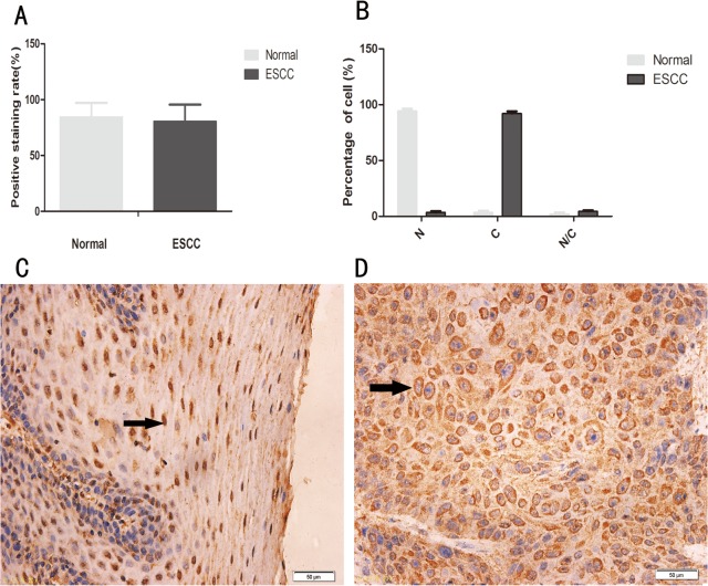
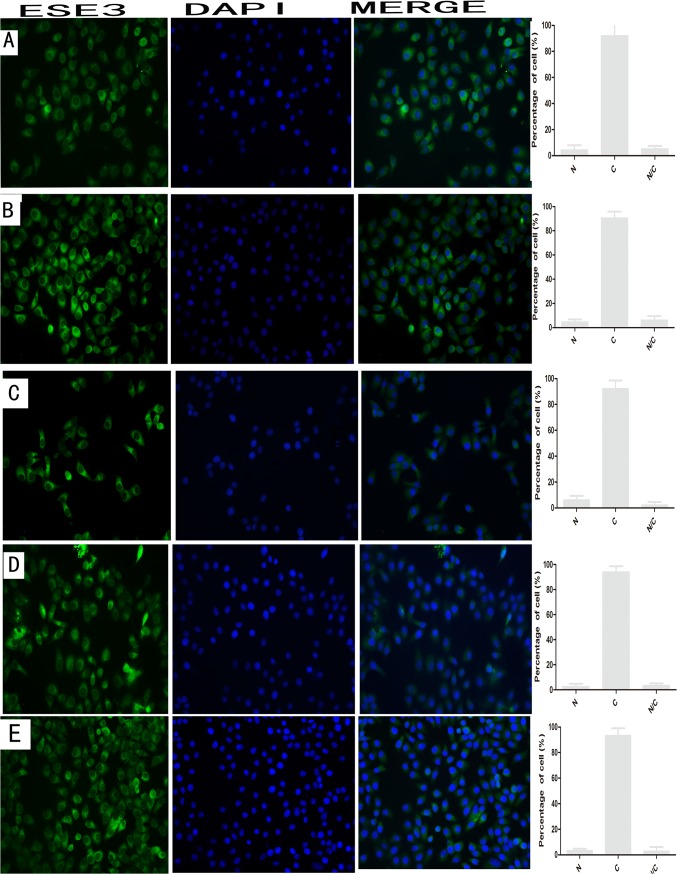
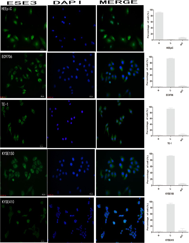
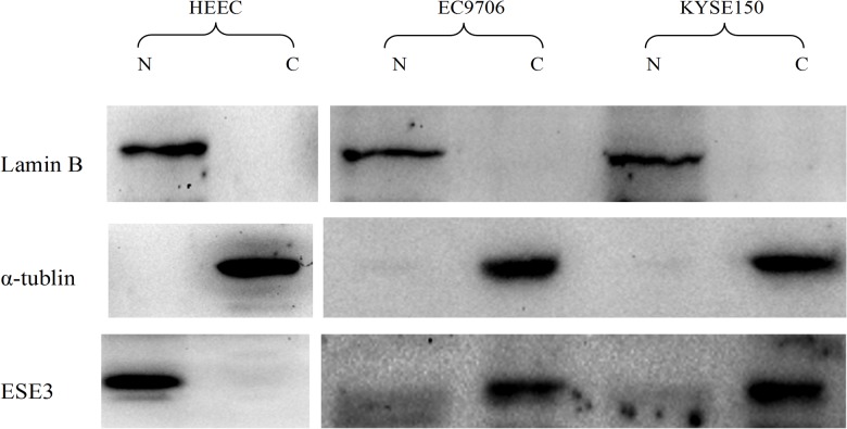
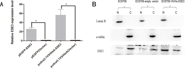
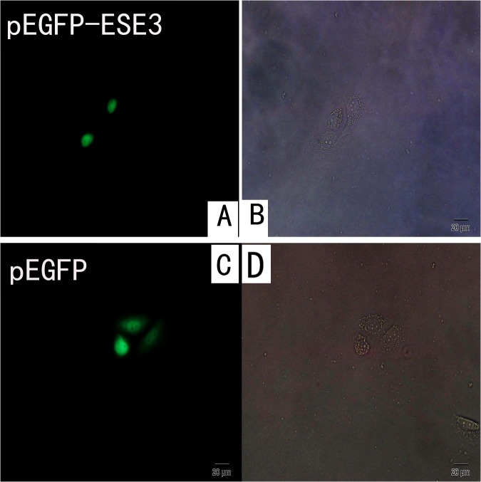
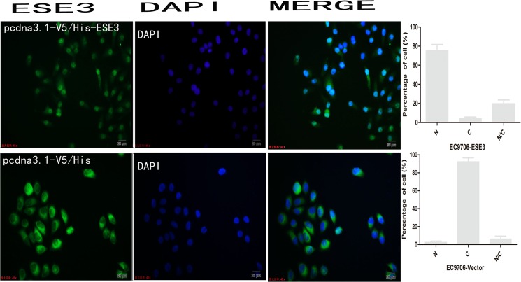
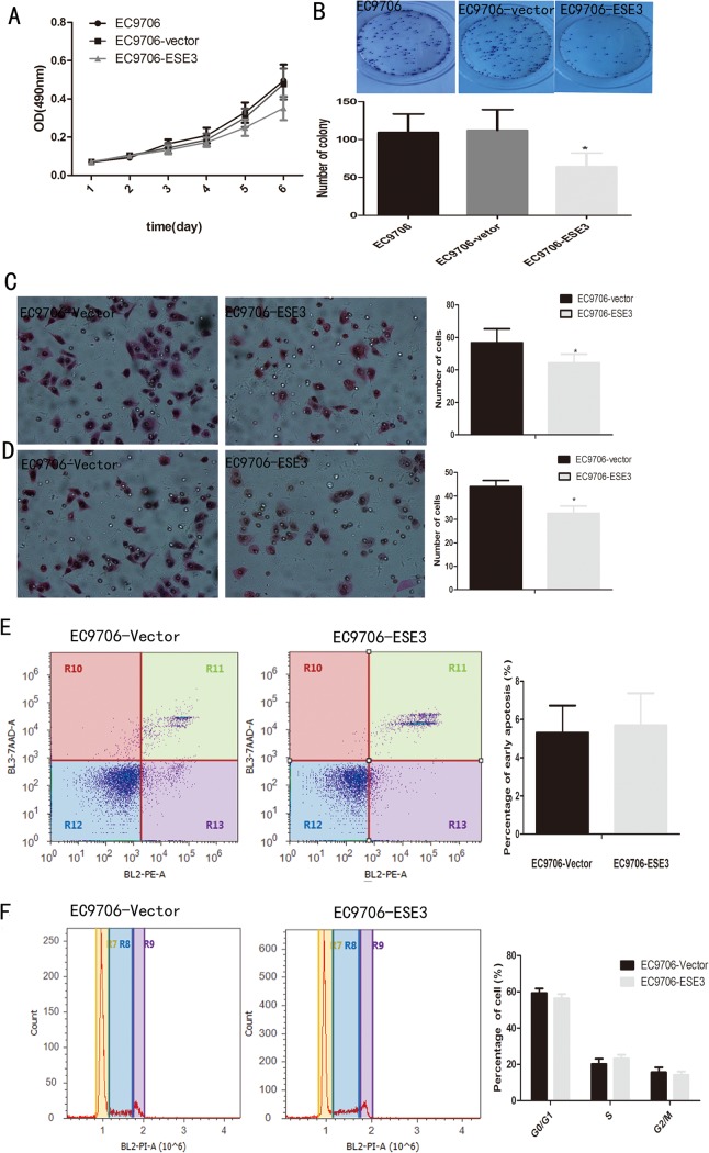
Esophageal cancer is one of the most common malignant cancers worldwide. The molecular mechanism of esophageal squamous cell carcinoma (ESCC) is still poorly understood. ESE3 is a member of the Ets transcription family, which is only expressed in epithelial tissues and acts as a tumor suppressor gene in prostate cancer. Our study aim was to confirm whether ESE3 is involved in the carcinogenesis of ESCC. Immunohistochemical analysis revealed that ESE3 was mainly located in cell nuclei of normal tissues and the cytoplasm in ESCC tissues. Immunofluorescence and western blot analyses of the normal esophageal cell line HEEpiC and ESCC cell lines EC9706 TE-1, KYSE150, and KYSE410 confirmed these results. pEGFP-ESE3 and pcDNA3.1-V5/HisA-ESE3 plasmids were constructed for overexpression of ESE3 in EC9706 and KYSE150 cells. The stably transfected cells showed restoration of the nuclear localization of ESE3. EC9706 cells with re-localization of ESE3 to the nucleus showed inhibition of proliferation, colony formation, migration, and invasion. To explore the possible mechanism of the differences in localization of ESE3 in normal esophageal cells and ESCC cells, ESCC cell lines were treated with the nuclear export inhibitor leptomycin B, transcription inhibitor actinomycin D, PKC inhibitor sphinganine, P38 MAPK inhibitor SB202190, and CK II inhibitor TBCA. These reagents were chosen according to the well-known mechanisms of protein translocation. However, the localization of ESE3 was unchanged after these treatments. The sequence of ESE3 cDNA in ESCC cells was identical to the standard sequence of ESE3 in the NCBI Genebank database, indicating that there was no mutation in the coding region of ESE3 in ESCC. Taken together, our study suggests that ESE3 plays an important role in the carcinogenesis of ESCC through changes in subcellular localization and may act as a tumor suppressor gene in ESCC, although the mechanisms require further study.
食管癌是全球最常见的恶性肿瘤之一。食管鳞状细胞癌(ESCC)的分子机制仍未完全清楚。ESE3是Ets转录家族的成员之一,仅在上皮组织中表达,在前列腺癌中作为肿瘤抑制基因发挥作用。我们的研究目的是确认ESE3是否参与ESCC的致癌过程。免疫组织化学分析显示,ESE3主要位于正常组织的细胞核中,而在ESCC组织中位于细胞质中。对正常食管细胞系HEEpiC和ESCC细胞系EC9706、TE-1、KYSE150和KYSE410进行免疫荧光和蛋白质印迹分析证实了这些结果。构建了pEGFP-ESE3和pcDNA3.1-V5/HisA-ESE3质粒,用于在EC9706和KYSE150细胞中过表达ESE3。稳定转染的细胞显示ESE3的核定位得以恢复。ESE3重新定位于细胞核的EC9706细胞表现出增殖、集落形成、迁移和侵袭受到抑制。为了探究ESE3在正常食管细胞和ESCC细胞中定位差异的可能机制,用核输出抑制剂放线菌素B、转录抑制剂放线菌素D、蛋白激酶C抑制剂鞘氨醇、P38丝裂原活化蛋白激酶抑制剂SB202190和酪蛋白激酶II抑制剂TBCA处理ESCC细胞系。这些试剂是根据已知的蛋白质转运机制选择的。然而,这些处理后ESE3的定位没有改变。ESCC细胞中ESE3 cDNA的序列与NCBI基因库数据库中ESE3的标准序列相同,表明ESCC中ESE3的编码区没有突变。综上所述,我们的研究表明ESE3通过亚细胞定位的改变在ESCC的致癌过程中发挥重要作用,并且在ESCC中可能作为肿瘤抑制基因,尽管其机制需要进一步研究。