Bomsztyk Karol, Mar Daniel, An Dowon, Sharifian Roya, Mikula Michal, Gharib Sina A, Altemeier William A, Liles W Conrad, Denisenko Oleg
UW Medicine South Lake Union, University of Washington, 850 Republican Street, 98109, Seattle, WA, USA.
Department of Medicine, University of Washington, 850 Republican Street, 98195, Seattle, WA, USA.
Crit Care. 2015 May 11;19(1):225. doi: 10.1186/s13054-015-0943-4.
The Tie2/angiopoietin (Tie2/Ang) and vascular endothelial growth factor receptor-ligand systems (VEGFR/VEGF) are recognized to play important roles in the regulation of microvascular endothelial function. Downregulation of these genes during sepsis has been implicated in the pathogenesis of sepsis-related microvascular leak and multiple organ dysfunction syndrome. Mechanisms responsible for dysregulation of angiogenic genes in sepsis are poorly defined.
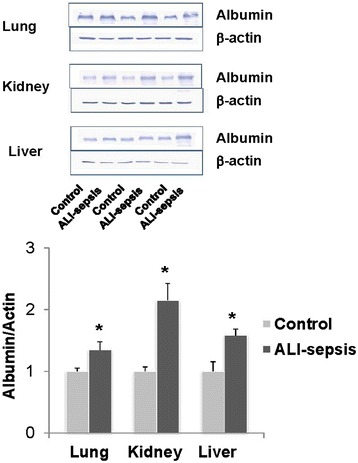
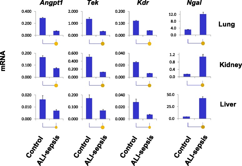
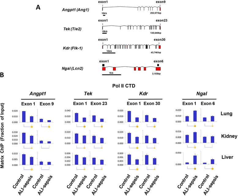
Western blot, reverse transcription-polymerase chain reaction, and multiplex chromatin immunoprecipitation platform (Matrix ChIP) were used to investigate serum albumin leak, changes in gene expression, and associated epigenetic alterations in a murine model of acute lung injury-induced sepsis (ALI-sepsis).
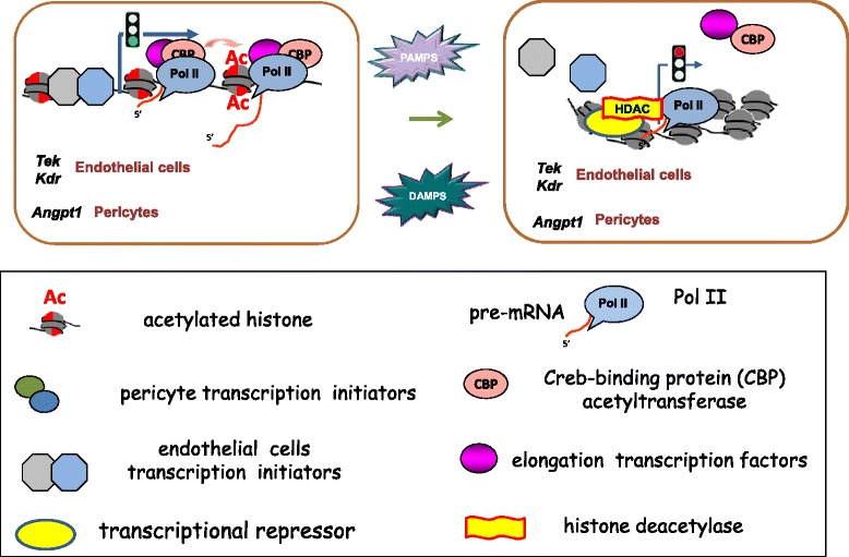
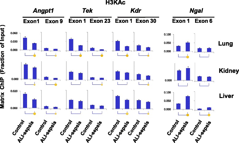
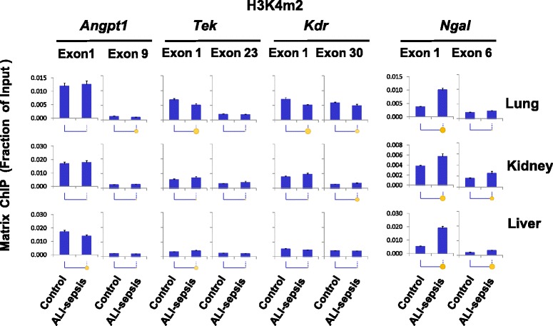
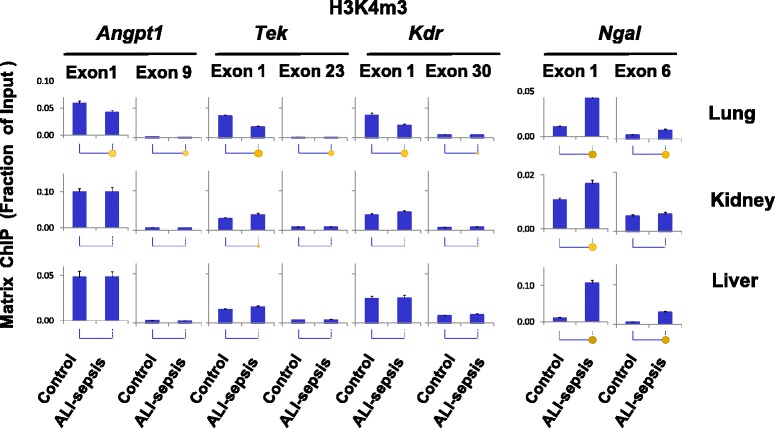
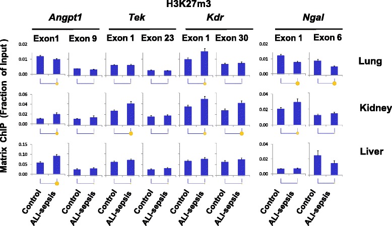
Experimental ALI-sepsis induced microvascular leak and downregulation of expression of Angpt1 (Ang1), Tek (Tie2), and Kdr (Vegfr2 or Flk-1) genes in the lung, kidney, and liver. These changes correlate with a decrease in RNA polymerase II density at these genes, and the greatest response was observed in the lung. ALI-sepsis reduced levels of transcription-permissive histone H3 lysine acetylation (H3KAc) at these loci in all examined tissues. Decreases in permissive H3K4m3 and H3Km2 marks were detected only in the lung. In contrast, only minimal alterations in transcription-repressive histone modifications (H3K27m3, H3K9m2, H3K9m3, and H4K20m3) were observed in all tissues.
Our results demonstrate that decreases in transcription-permissive, but not increases in transcription-repressive, histone modifications at Angpt1, Tek, and Kdr are a systemic, rather than a lung-restricted, response, involving key end-organs in experimental ALI-sepsis. Given that ventilator-associated pneumonia is a major cause of sepsis in critically ill patients, elucidation of mechanisms mediating epigenetic alterations during sepsis provides fundamental new insights into the pathogenesis of sepsis-induced microvascular leak and subsequent end-organ injury/dysfunction.
Tie2/血管生成素(Tie2/Ang)和血管内皮生长因子受体-配体系统(VEGFR/VEGF)被认为在微血管内皮功能调节中发挥重要作用。脓毒症期间这些基因的下调与脓毒症相关微血管渗漏和多器官功能障碍综合征的发病机制有关。脓毒症中血管生成基因失调的机制尚不清楚。
采用蛋白质免疫印迹法、逆转录-聚合酶链反应和多重染色质免疫沉淀平台(Matrix ChIP),研究急性肺损伤诱导的脓毒症(ALI-脓毒症)小鼠模型中的血清白蛋白渗漏、基因表达变化及相关表观遗传改变。
实验性ALI-脓毒症诱导了肺、肾和肝脏中的微血管渗漏以及血管生成素1(Ang1)、Tek(Tie2)和激酶插入域受体(Kdr,即血管内皮生长因子受体2或Flk-1)基因表达下调。这些变化与这些基因处RNA聚合酶II密度降低相关,且在肺中观察到的反应最为明显。ALI-脓毒症降低了所有检测组织中这些基因座处的转录许可组蛋白H3赖氨酸乙酰化(H3KAc)水平。仅在肺中检测到转录许可组蛋白H3赖氨酸4三甲基化(H3K4m3)和组蛋白H3赖氨酸79二甲基化(H3Km2)标记减少。相比之下,在所有组织中仅观察到转录抑制组蛋白修饰(H3K27m3、H3K9m2、H3K9m3和H4K20m3)的微小改变。
我们的结果表明,在血管生成素1、Tek和激酶插入域受体基因处,转录许可组蛋白修饰的减少而非转录抑制组蛋白修饰的增加是一种全身性反应,而非仅限于肺部的反应,涉及实验性ALI-脓毒症中的关键终末器官。鉴于呼吸机相关性肺炎是重症患者脓毒症的主要原因,阐明脓毒症期间介导表观遗传改变的机制可为脓毒症诱导的微血管渗漏及随后的终末器官损伤/功能障碍的发病机制提供全新的重要见解。